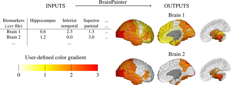
Figure 1:
Given a .csv file with region-of-interest (ROI) biomarkers and a user-defined color gradient, BrainPainter can automatically generate brain images with the cortical surface (left and middle) as well as with subcortical structures (right). The input .csv file can have multiple rows, one for each set of output images. The color gradient is a list of RGB colours given by the user. Final colours are interpolated using the numbers from the input .csv file based on the color gradient – e.g. if the hippocampus has a value of 1.2, its final color will be an interpolation of colors 1 (yellow) and 2 (orange) from the gradient.

