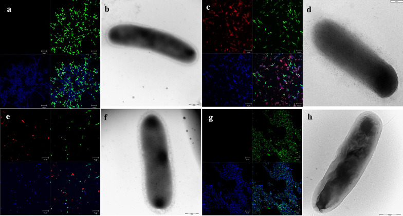
Fig. 6.
Confocal and Transmission electron microscopy of BamE expressed in E. coli. E. coli BL21(DE3) expressing the full-length neisserial BamE (a and b), or fused to AIDA-I (c and d), or the E. coli BamE fused to Lpp’OmpA (e and f) grown at 25 °C, were incubated first with anti-FLAG antibodies while E. coli T7ExpressIq (pET15b) expressing InaK fused to the neisserial BamE grown at 18 °C, was incubated first with polyclonal anti-NmBamE antibodies (g and h). Subsequently samples were incubated with the secondary anti-mouse immunoglobulin G (whole molecule) Alexa fluor 568-conjugated. The lipoproteins can be visualized in red, the DNA in blue (DAPI) and the membranes in green (oregon green) (a, c, e and f). In transmission electron microscopy using immunogold labelling, the same samples were incubated with the secondary anti-mouse immunoglobulin G conjugated with 5 nm gold particles (b, d, f and h)

