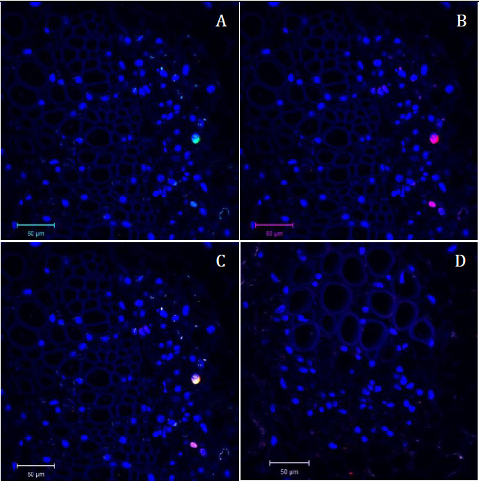
Figure 4. Confocal microscopy pictures of double labeling (FISH/Immuno) experiment obtained following our protocol.

Cross sections of infected faba bean petioles stained with DAPI (in blue) and hybridized with the S-A488 probe in green and M-Rep protein antibody in red. A. Green channel (S probe = part of the segment encoding the capsid protein) merged with the blue channel (DAPI). B. Red channel (M-Rep protein localization = immunolabeling of the movement protein) merged with the blue channel (DAPI). C. Merged image of the three channels. D. Merged picture of the three channels of the control sample (petiole of non-infected faba bean plant) taken in the same conditions as Figure 4C; only autofluorescence of the tissue and blue signal of the DAPI nuclei staining could be observed.
