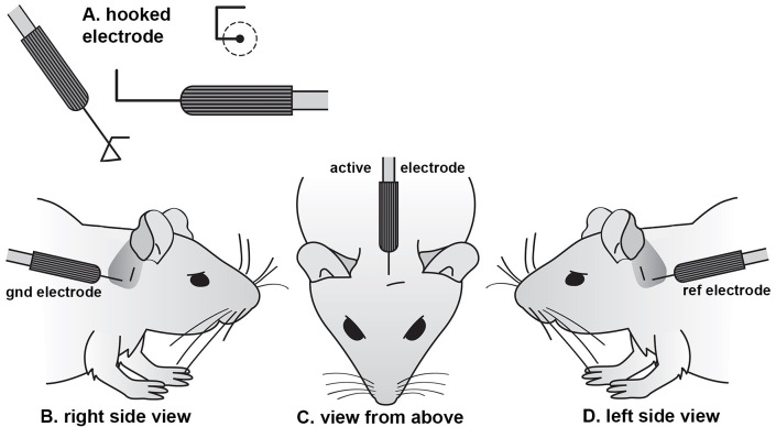
Figure 1. Subcutaneous electrode shape and placement.
(A) Individual needle electrodes are bent into a square hook shape using a needle holder, with each side approxiamtely 2 mm long. The hook is inserted through a pinch of skin, such that the contact with the subdermal layers lies on the remaining straight shaft of the electrode. A ground electrode is placed in the skin overlying the bulla behind the right ear (B). The active electrode for the low-impedance bio-amplifier is placed on the midline in a rostro-caudal position lined up with the leading edge of the 2 pinnae (C). The reference electrode for the bio-amplifier is inserted behind the left ear (D), in an equivalent location to the ground electrode (modified from Ingham et al., 2011 ).

