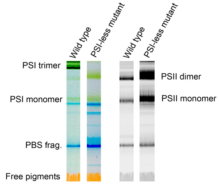
Figure 2. Representative color scans (left pair) and chlorophyll fluorescence images (right pair) of the gel with separated Photosystem I and Photosystem II complexes from the cyanobacterium Synechocystis PCC 6803 GT-P .
( Tichý et al., 2016 ) and the Photosystem I-less mutant ( Shen et al., 1993 ). Left two lanes show color scans, right ones Chl fluorescence scans with dominant bands of Photosystem II dimer and monomer. Blue bands (PBS frag.) represent fragments of phycobilisomes, the cyanobacterial antennae, fluorescence of which is not elicited by blue light.

