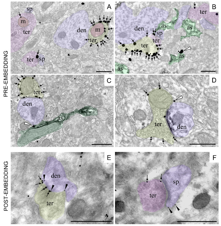
Figure 3. CB1 receptor immunolocalization in different subcellular compartments of the rodent brain.
Single pre-embedding immunogold (A) and double pre-embedding immunogold and immunoperoxidase methods (B-D). A. CB1 receptor labeling (arrows) at a presynaptic GABAergic terminal (ter, yellow) adjacent to a dendrite (den, purple). CB1 receptor particle is localized to a presynaptic glutamatergic terminal (ter, pink) associated with a spine (sp, purple). Mitochondria (m, red) exhibit CB1 receptor immunolabeling in both glutamatergic (ter, pink) and GABAergic (ter, yellow) presynaptic terminals (CA1 stratum radiatum, adult mouse hippocampus). B. CB1 receptor labeling (arrows) at a presynaptic GABAergic terminal (ter, yellow), glutamatergic terminals (ter, purple) and in one astrocyte branch (white arrowhead; as, green) in the mouse piriform cortex. Astrocytes are labeled with anti-GLAST/immunoperoxidase/DAB method (black precipitate in as). C. CB1 receptor labeling (arrows) at a presynaptic GABAergic terminal (ter, yellow) adjacent to a dendrite (den, purple) and in one astrocyte process (white arrowhead; as, green) in the molecular layer of the mouse dentate gyrus. Astrocytes are labeled with anti-GFAP/immunoperoxidase/DAB method (black precipitate in as). D. CB1 receptor labeling (arrows) at a presynaptic terminal (ter, yellow) combined with anti-gephyrin/immunoperoxidase/DAB method (black precipitate in den, purple) to positively identify the inhibitory postsynaptic membrane of a GABAergic synapse. White arrowhead: CB1 receptor labeling at a thin astrocytic process filling the intercellular space (rat prelimbic cortex). E-F. Double post-embedding immunogold method revealing the localization of presynaptic CB1 receptors (ter; 18 nm-diameter gold particles) and postsynaptic mGluR2/3 (den, sp; 10 nm-diameter gold particles) at inhibitory (ter, yellow) and excitatory (ter, pink) synapses in the mouse dentate molecular layer. Scale bars= 500 nm.

