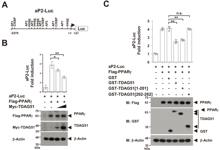
Fig. 3. TDAG51 inhibits PPARγ-induced aP2 promoter activity.
(A) Schematic diagram of the aP2 promoter (aP2-Luc). The putative transcription factor binding sites are illustrated, and the nucleotide positions are numbered based on the transcriptional start site. PPRE, PPARγ responsive element; C/EBP, CCAAT/enhancer binding protein; SP1, specificity protein 1; AP-2, activator protein 2. (B) The inhibitory effect of TDAG51 on PPARγ-induced aP2-Luc reporter activity. Reporter plasmids (aP2-Luc [0.1 µg] and pcDNA3.1/His/LacZ [0.1 µg]) were cotransfected with epitope-tagged expression plasmids (Flag-PPARγ [0.3 µg] and/or Myc-TDAG51 [0.2 and 0.5 µg]) into 293T cells. The expression of transfected plasmids was analyzed by immunoblotting against anti-epitope antibodies. β-Actin was used as the loading control. Protein expression is indicated by an arrow. (C) The inhibitory effects of TDAG51 deletion mutants on PPARγ-induced aP2-Luc reporter activity. Reporter plasmids were cotransfected with epitope-tagged expression plasmids (Flag-PPARγ [0.3 µg] and/or GST-TDAG51 [0.5 µg]) into 293T cells. GST alone (mock) was used as a control. *P < 0.05; **P < 0.01; n.s., not significant.

