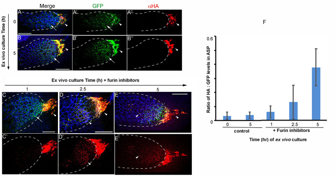
Figure 3. Ex vivo culture and Furin inhibitor assay on wing discs expressing Bnl:HA1GFP3.
The α-HA stained (red) wing discs that expressed Bnl:HA1GFP3 under bnl-Gal4 were ex vivo-cultured for 0 (pre-treatment) and 5 h in the absence (A-B") and 1, 2.5 and 5 h in the presence of Furin inhibitors (C-E') as indicated; arrow, truncated Bnl:GFP3 derivative; arrowhead, uncleaved Bnl:HA1GFP3; blue, α-Discs large to mark cell outlines; merged (A, B, C-E) and either split green, red (A’-B”) or only red (C’-E’) channels were shown. (F) Graph comparing average levels of colocalized HA and GFP in ASPs cultured in the presence and absence of Furin inhibitors; samples were harvested at different time points from the continuous culture; N = 11 (0 h), 11 (1 h), 10 (2.5 h), 9 (5 h control), 12 (5 h test); P-values (ANOVA followed by Tukey HSD): P = 0.0001 for 5 h vs. either 0 h control, 5 h control, 1 h, or 2.5 h. Scale bars, 30 μm.

