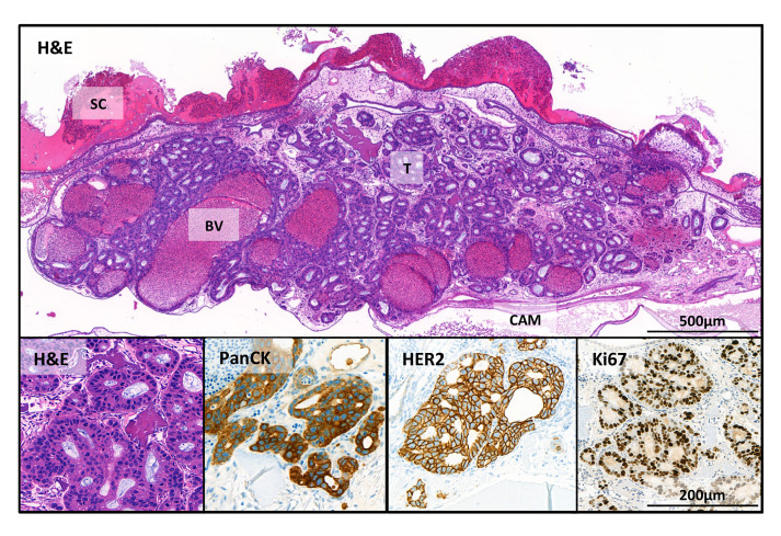
Figure 4. Immunohistochemical stains of the tumor in the CAM.
OE19 esophageal adenocarcinoma cells (1 x 10⁶ inoculated on CAM for 7 days). SC = scaffold; BV = blood vessels; T = tumor and CAM = chorioallantoic membrane. Shown here are stains with hematoxylin and eosin (H&E); and human specific IHC stains: pan-cytokeratin (PanCK), an epithelial marker; HER2, a receptor tyrosine kinase, which is amplified in OE19 cells and Ki-67 that is associated with proliferation.

