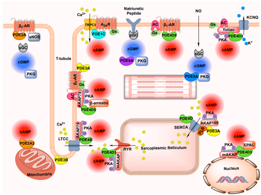
Figure 1: Schematic representation of cyclic nucleotide and PDE compartmentalization in cardiac myocyte.

Compartmentalized cyclic nucleotide signaling is generated by individual G-protein coupled receptors, and established via the formation of multiple spatially segregated signalosomes, in which combination of cyclic nucleotide effectors, phosphodiesterase and scaffolding proteins form specific complexes via protein-protein interaction. PDE, phosphodiesterase; AR, adrenergic receptor; AC, adenylyl cyclase; sGC, soluble guanylyl cyclase; pGC, particulate guanylyl cyclase; PKA, protein kinase A; PKG, protein kinase G; cAMP, cyclic adenosine 3′,5′-monophosphate; cGMP, cyclic guanosine 3′,5′-monophosphate; LTCC, L-type calcium current; PLB, phospholamban; RYR, ryanodine receptor; SERCA, sarcoplasmic reticulum Ca2+ ATPase; AKAP, A-kinase anchor protein; TRPC, transient receptor potential subfamily C; A2AR, adenosine type 2A receptor; KCNQ, voltage-gated potassium channels of the KCNQ subfamily; NO, nitric oxide; EPAC, exchange protein activated by cAMP; eNOS, endothelial nitric oxide synthase.
