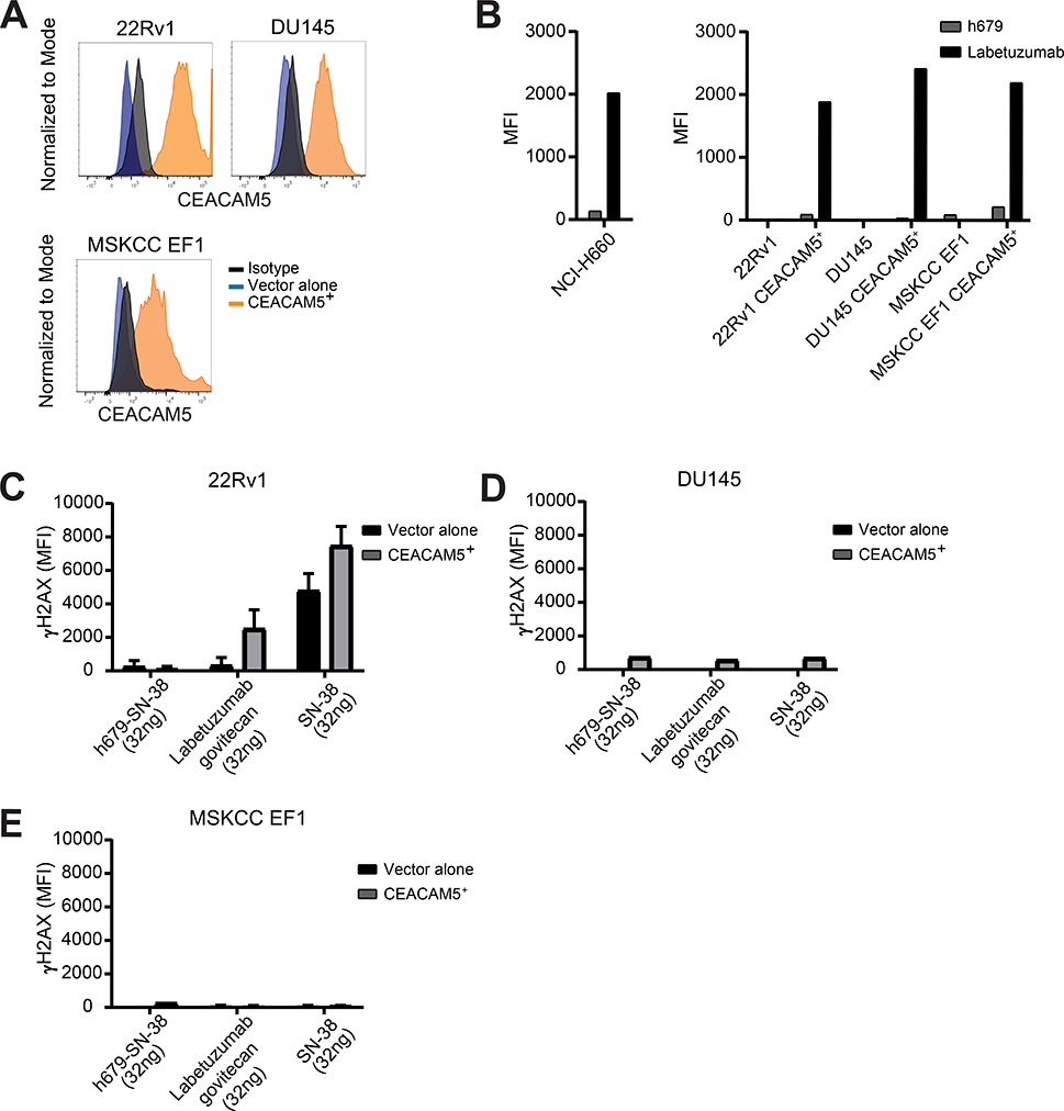
Figure 5. Labetuzumab govitecan induces dsDNA damage in a CEACAM5-specific manner.
(A) CEACAM5 surface protein expression determined by flow cytometry in prostate cancer cell lines transduced with lentiviral expression constructs. (B) Labetuzumab binding to CEACAM5 in prostate cancer cell lines. Measurement of intracellular γH2AX staining of (C) 22Rv1, (D) DU145, and (E) MSKCC EF1 cells 16 hours after treatment with h679-SN-38, labetuzumab govitecan, or SN-38 for 30 minutes on ice. MFI = Mean fluorescence intensity. Histograms depict means + SD for experimental duplicates.

