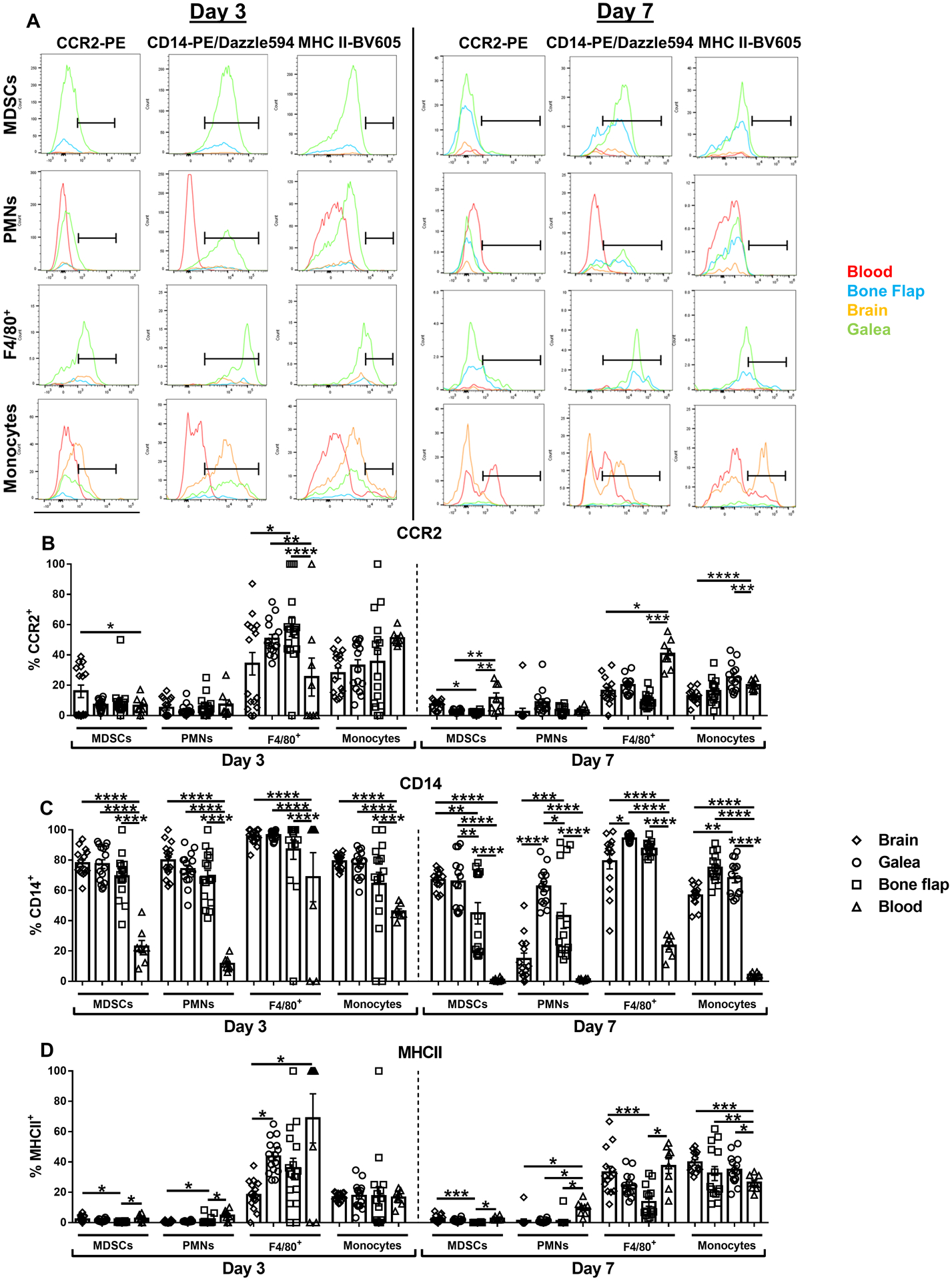
Fig. 2. Differential expression of leukocyte markers in tissue compartments during S. aureus craniotomy infection.

Mice were sacrificed at days 3 (n=16) or 7 (n=14) following S. aureus craniotomy infection, whereupon CCR2, CD14, and MHC Class II expression was quantified by flow cytometry. (A) Representative histograms of leukocyte populations in the brain, galea, bone flap, and blood are shown and the percentage of viable leukocytes expressing (B) CCR2, (C) CD14, and (D) MHC Class II are shown. Results represent the mean ± SEM combined from two independent experiments and analyzed by One-way ANOVA with Tukey’s multiple comparison test (*, p < 0.05; **, p < 0.01; ***, p < 0.001; ****, p < 0.0001).
