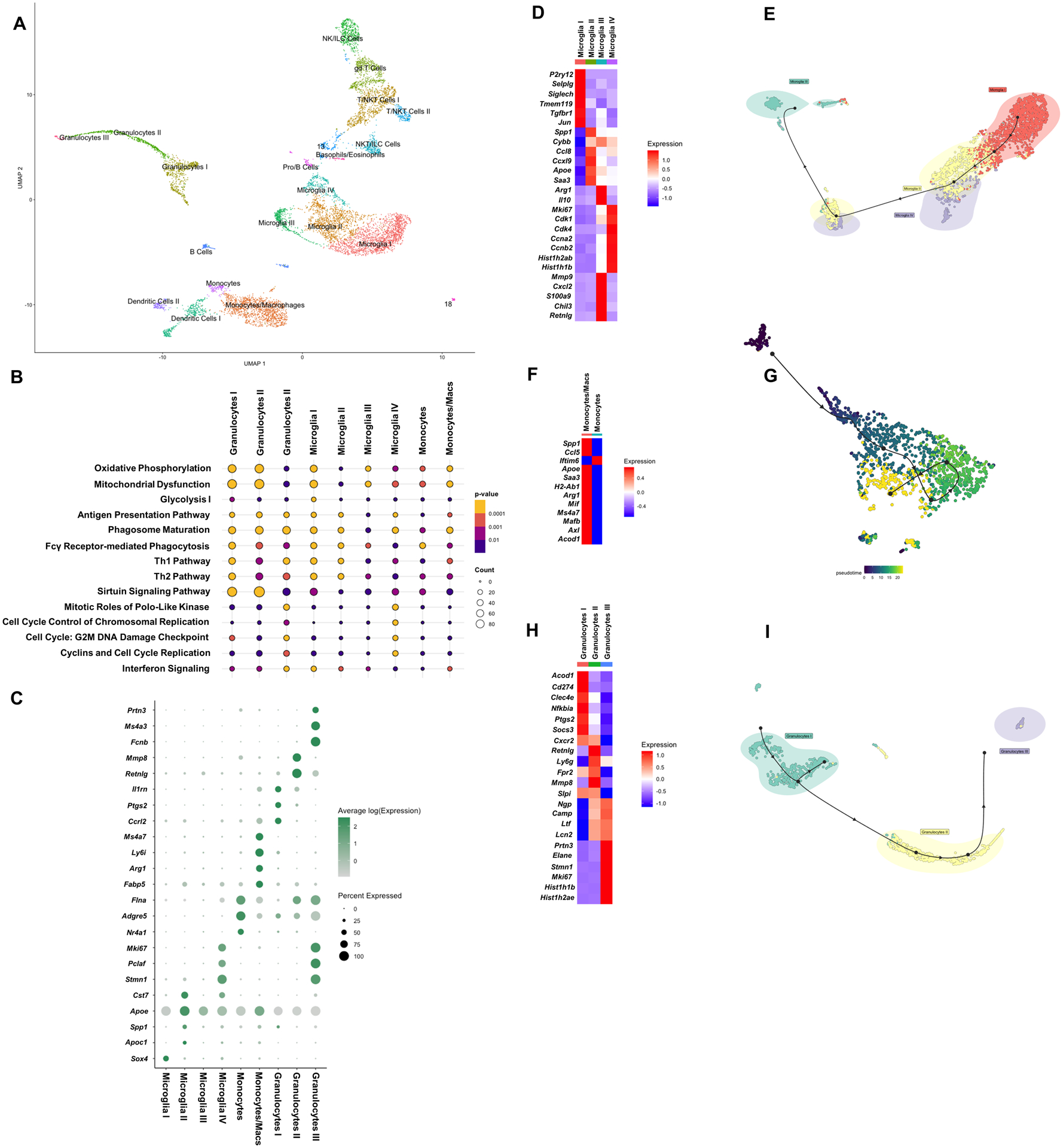
Fig. 5. Transcriptional heterogeneity in microglia and innate immune cells in the brain during S. aureus craniotomy infection.

Viable CD45+ cells were isolated from the brain (n= 9,233 cells) of mice (n=10) at day 7 following S. aureus craniotomy infection by FACS and processed for scRNA-seq. (A) Transcriptional clusters were identified by SingleR and are presented as UMAP plots. (B) Ingenuity Pathway Analysis (IPA) of canonical pathways that were significantly expressed in microglia and other brain leukocyte infiltrates and (C) genes that were enriched in specific myeloid clusters in the brain (p < 0.05). Heatmaps and trajectory analysis of (D and E) microglia, (F and G) monocyte/macrophage, and (H and I) granulocyte clusters.
