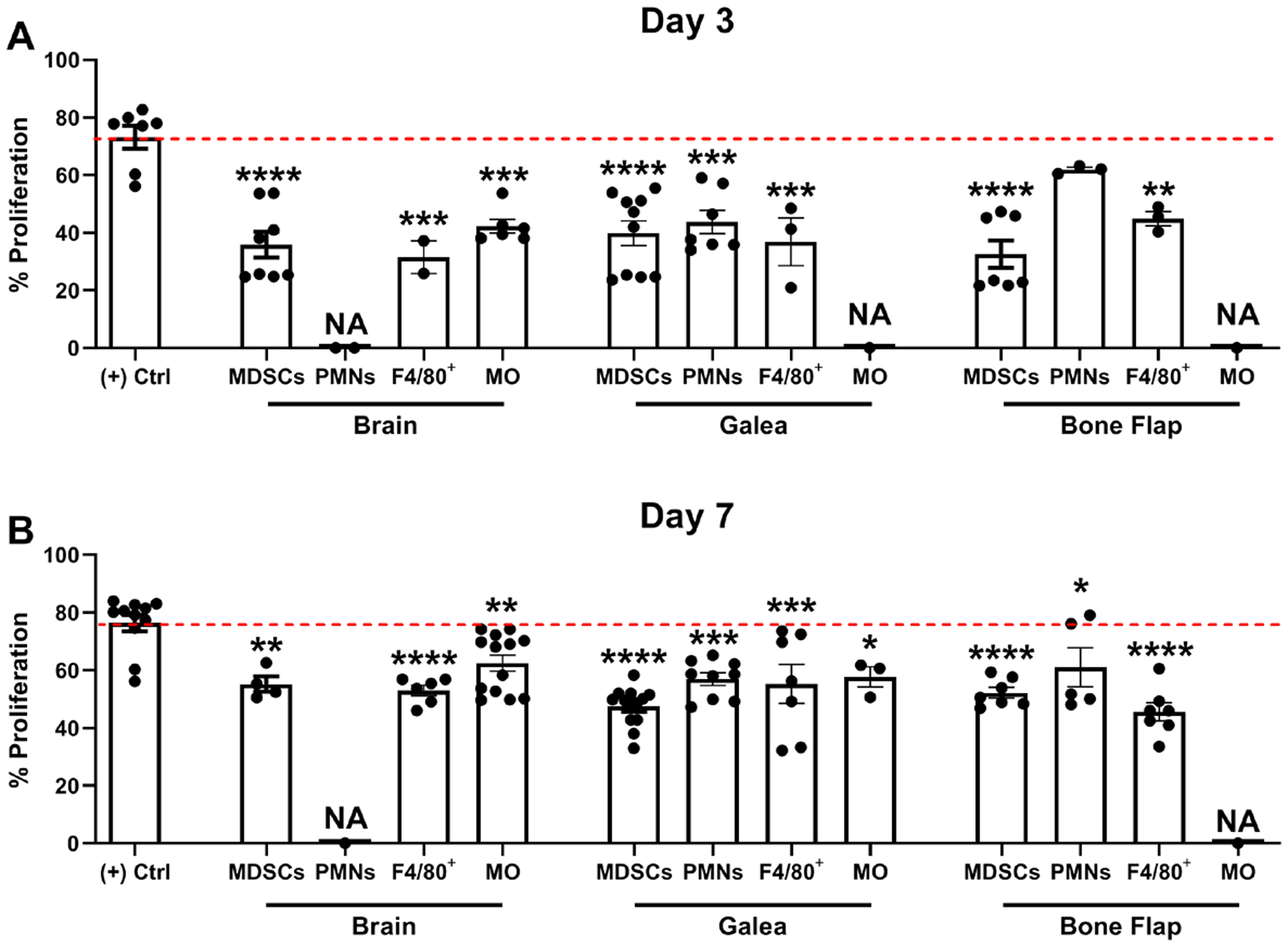
Fig. 8. Leukocytes recovered from the brain, bone flap, and galea inhibit T cell proliferation.

Leukocyte populations were isolated from the brain, galea, or bone flap at (A) 3 or (B) 7 days following S. aureus craniotomy infection by FACS and assessed for their ability to inhibit CD4+ T cell proliferation at a 1:1 ratio (T cell:MDSC, F4/80+, PMN, or monocyte; MO). Results are expressed as the percentage of proliferation compared to CD3/CD28-stimulated T cells (+ Control (Ctrl); red dotted line). A sufficient number of leukocytes could not be recovered from various compartments for this assay (NA, not applicable). Results represent the mean ± SD combined from three independent experiments and analyzed by One-way ANOVA with Tukey’s multiple comparison test (*, p < 0.05; **, p < 0.01; ***, p < 0.001; ****, p < 0.0001 compared to the (+) control).
