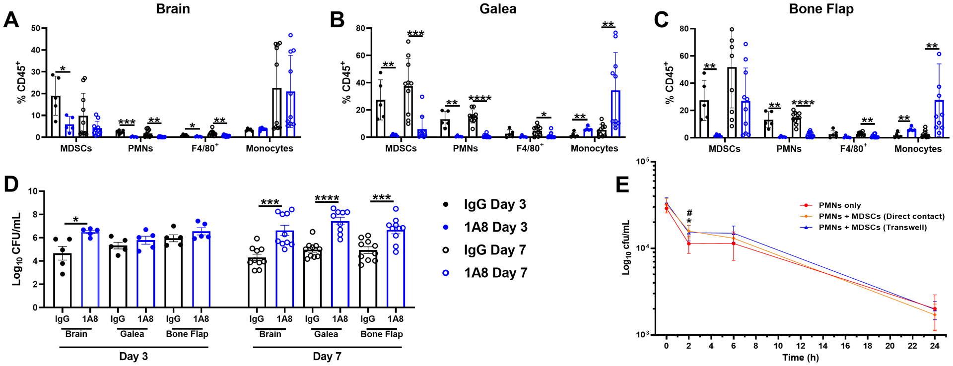
Fig. 9. Ly6G+ PMNs are critical for bacterial containment during S. aureus craniotomy infection.

Mice received anti-Ly6G (1A8) or isotype-matched control antibody (100 μg/mouse) beginning one day prior to S. aureus craniotomy infection and every two days until animals were sacrificed. Leukocyte infiltrates in the (A) brain, (B) galea, and (C) bone flap were quantified by flow cytometry with (D) bacterial burden in each compartment determined. Results represent the mean ± SD from one (day 3; n=5 mice/group) or two combined independent experiments (day 7; n=10 mice/group) and analyzed by One-way ANOVA with Tukey’s multiple comparison test (*, p < 0.05; **, p < 0.01; ***, p < 0.001; ****, p < 0.0001). (E) PMNs were exposed to live S. aureus USA300 LAC for 2 h at an MOI of 10:1 (bacteria:cell) in the presence or absence of MDSCs in direct contact or separated by Transwell inserts to evaluate the effects on PMN S. aureus killing by gentamicin protection assays. Results are combined from two independent experiments (n = 5–8 biological replicates) and are presented as the mean ± SD. (*, p < 0.05 for PMNs only vs. PMNs + MDSCs in direct contact; #, p < 0.05 for PMNs only vs. PMNs + MDSCs with Transwells; One-way ANOVA with Tukey’s multiple comparisons test).
