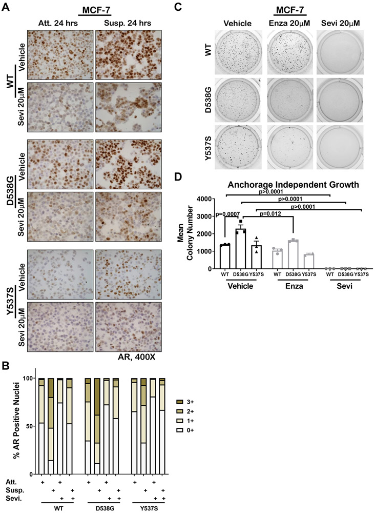
Figure 3. AR increased in breast cancer cells grown in soft agar and AR inhibition abolished the selective advantage of mutant ER cells for anchorage-independent survival.
A. WT, D538G, and Y537S mutant MCF7 cells were grown in full serum media under attached (Att) or suspension conditions (on poly-HEMA plates, Susp) and treated ± 20μM Seviteronel (Sevi) for 24 hours. Cells were pelleted, FFPE, and AR IHC performed. Representative images are shown at 400x. B. Quantification of A with ImageScope software for percent positive nuclei for staining intensities of 0-3+ for all conditions attached (Att), suspended (Susp), and ± Sevi. C. WT, D538G, and Y537S cells plated in 0.3% agar and treated with 20μM Sevi, 20μM enzalutamide (Enza) or EtOH control and grown for three weeks with bi-weekly media and drug changes. Representative images of anchorage-independent growth are shown. D. Colony number quantified using ImageJ software for assay in C (N=3). Mean ± SEM, One way ANOVA with Tukey’s multiple comparison test is depicted. Two way ANOVA, interaction between cell line and treatment p = 0.003.

