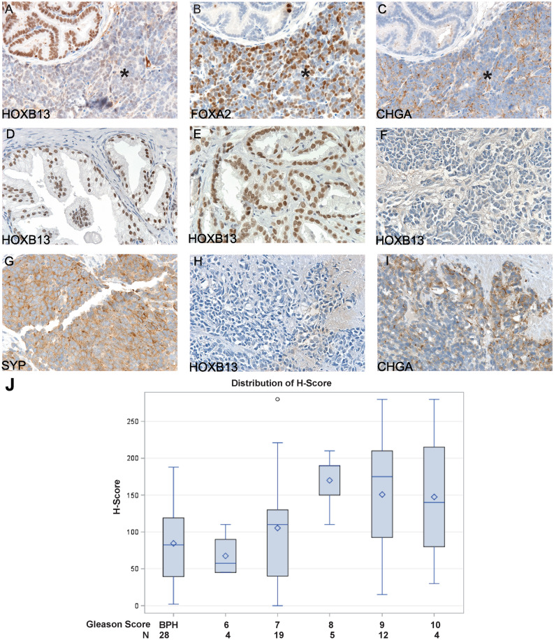
Figure 3.
Immunohistochemical staining to evaluate the expression of HOXB13 in prostatic tissues. (A–C) Serial sections of a TRAMP NEPCa tumor displayed low HOXB13 expression in NEPCa area (indicated by * sign) in contrast to the positive staining in focal PIN. NEPCa markers Chromogranin A (CHGA) and FOXA2 were stained positive in NEPCa cells but negative in PIN. (D) HOXB13 expression in BPH, n = 28. (E) HOXB13 expression in AdPCa, n = 44. (F–I) Human NEPCa tumors demonstrated reduced (n = 1) or negative (n = 8) staining of HOXB13. These NEPCa tumors were stained positive for Synaptophysin (SYP) or Chromogranin A (CHGA). (J) Quantification of HOXB13 immunohistochemical staining with H-scores in benign prostatic hyperplasia (BPH) and prostatic acinar adenocarcinomas with a Gleason score of 6 (3 + 3), 7 (3 + 4 and 4 + 3), 8 (4 + 4), 9 (4 + 5 and 5 + 4), and 10 (5 + 5). The H score of HOXB13 immunohistochemical staining was correlated with Gleason score, Spearman correlation test, Rho = 0.348, p < 0.05.

