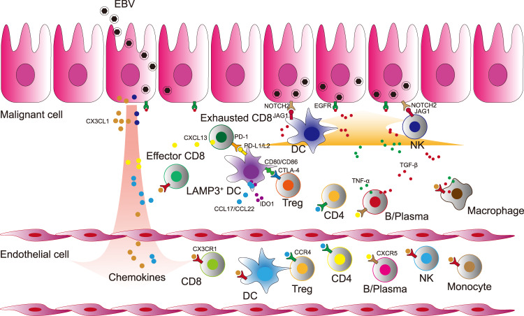
Fig. 7. Schematic diagram of cross-talks among multiple immune cells in the TME of NPC.
EBV infects nasopharyngeal epithelial cells and participates in the tumorigenic process of NPC. EBV-positive malignant NPC cells secret a variety of chemokines (CX3CL1, etc.) and initiate the recruitment and tumoral infiltration of multiple immune cells with the chemokines receptors from the peripheral blood. Multiple tumour infiltrating immune cells activate EGFR and Notch pathway in EBV-positive malignant NPC cells. Naive CD8+ cells infiltrate to the lesion and develop to effector and further exhausted CD8+ cells. Peripheral DCs infiltrate to the tumour and differentiate into LAMP3+ DCs. The mature LAMP3+ DCs with the expression of PD-L1/PD-L2 interact with PD1 on CD8+ T cells whereby the signalling restrains the activation of CD8+ T cells and promotes their exhaustion. Treg cells interact with LAMP3+ DCs through CTLA4-CD80/CD86, which might limit the antigen presentation process of DCs and promote the secretion of IDO1 to induce the proliferation of Treg cells. The intensive cell-cell interactions among LAMP3+ DCs, Treg cells, exhausted CD8+ T cells, and malignant cells foster an immune-suppressive niche for the tumour microenvironment of NPC.

