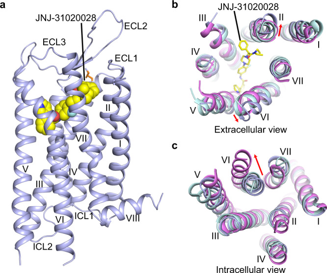
Fig. 1. Overall structure of Y2R–JNJ-31020028 complex.

a Side view of the Y2R–JNJ-31020028 structure. Y2R is shown in light blue cartoon representation. JNJ-31020028 (carbon in yellow, nitrogen in blue, oxygen in red, fluorine in cyan) is shown in sphere representation. The disulfide bond is shown as orange sticks. b, c Structural comparison of Y2R with inactive Y1R (PDB code: 5ZBQ) and active NTSR1 (PDB code: 6OS9). The helical bundles of Y2R, Y1R, and NTSR1 are colored light blue, light cyan, and pink, respectively. JNJ-31020028 is shown as sticks. b Extracellular view. Red arrows indicate the movements of helices II and VI in the Y2R structure compared to the structures of Y1R and NTSR1. c Intracellular view. Red arrow indicates the conformational change of helix VI in the active NTSR1 structure relative to the structures of Y1R and Y2R.
