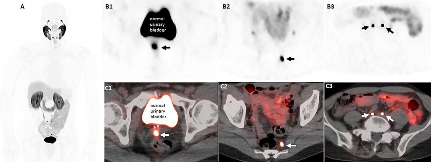
Figure 4:

64-years-old man with history of prostate cancer, Gleason 9 (4+5), status post EBRT and 1 year of ADT with PSA nadir of 0.4ng/ml, and recent rising PSA of 2.86ng/ml. 18F-DCFPYL PET/CT imaging, including maximal intensity projection (A), axial PET (B row) and axial fused PET/CT (C row) images demonstrate an intense DCFPYL avid focus at the midline of the seminal sevicles (B1, C1) and several subcentimeter pelvis lymph nodes including left presacral (B2, C2) and bilateral common iliac nodes (B2, C3).
