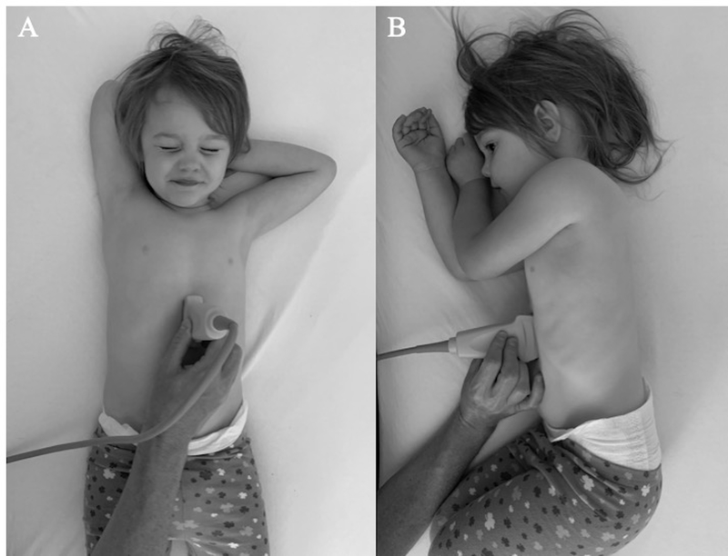
Figure 1.
Patient positioning and probe placement for gastric POCUS. A curvilinear or linear probe is placed in the midline sagittal plane inferior to the xiphoid process with the probe marker towards the patient’s head. Imaging is performed first in the supine position (A) and then the right lateral decubitus position (B).

