
Figure 1. Organization and structure of GPIb-IX.
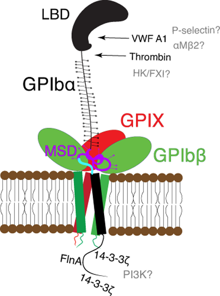
Cartoon illustration of the GPIb-IX complex including GPIbα (black), GPIbβ (green), GPIX (red). The N-terminal LBD of GPIbα is labeled, the membrane-proximal MSD is highlighted in purple, and the trigger sequence therein is highlighted in blue. The complex is held together by strong associations among the transmembrane domains as well as weak associations between GPIbβ and GPIX extracellular domains and potentially the MSD of GPIbα. Binding partners of the GPIbα LBD are listed to the right, including thrombin and VWF-A1. Intracellularly, 14-3-3ζ interacts with the intracellular tails of GPIbβ and GPIbα. FlnA binds to the tail of GPIbα. Binding partners for which a specific binding site has not been identified are listed in gray.