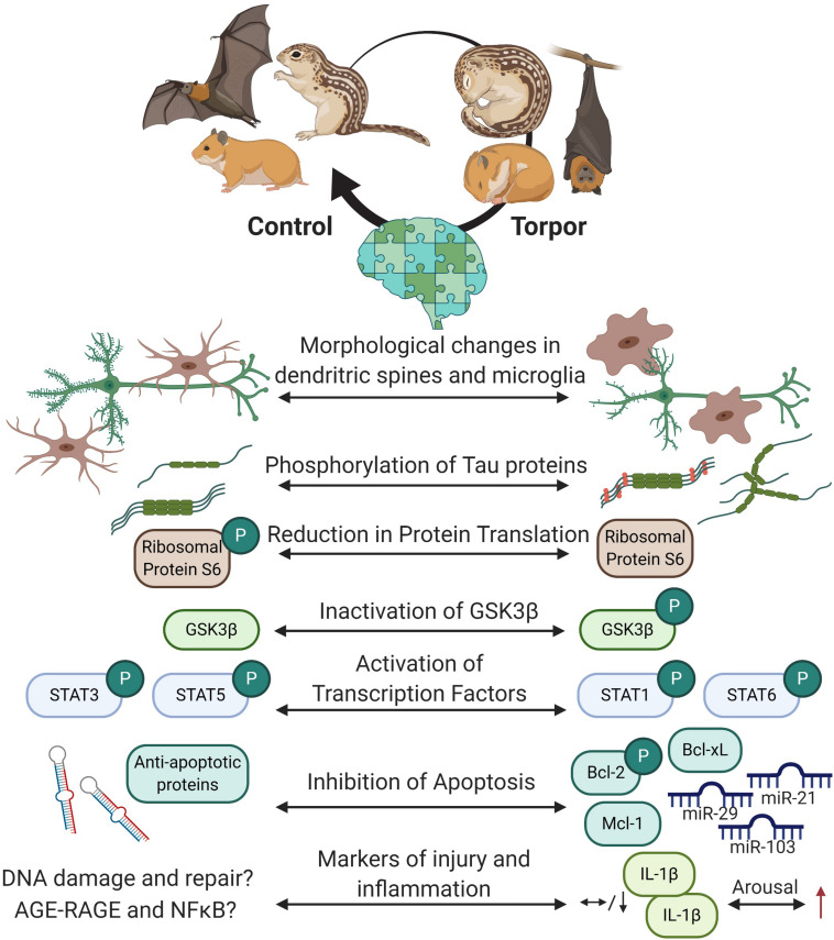
FIGURE 6.
Overview of the protective mechanisms in the hibernating brain. Cells within the hibernating brain undergo rapidly reversible morphological changes and changes to structural proteins to prevent cellular damage. Energy expensive processes like protein translation are inhibited. Transcription factors like such as signal transduction and activators of transcription (STAT) are differentially phosphorylated but not by glycogen synthase kinase 3 beta (GSK3β) during torpor, and may regulate apoptosis via B cell lymphoma proteins (Bcl-2 and Bcl-xL) or myeloid leukemia cell differentiation protein (Mcl-1) protein expression and phosphorylation. Some microRNAs are also increased in the brain to prevent cell death. Markers of brain injury and inflammation have yet to be fully characterized, but current research shows that some cytokines that are low in the brain during torpor increase during arousal. Created with BioRender.com.

