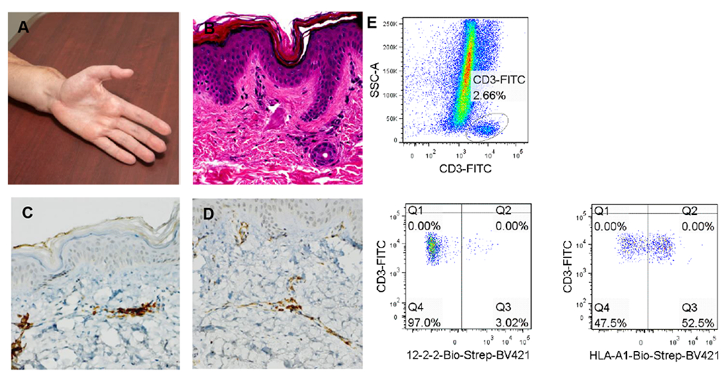
Figure 5: Recipient T cells identified in skin of transplanted human hand without evidence of rejection.

(A) Clinical photograph of transplant hand at 1 year. (B) H&E stained section of protocol biopsy obtained at 1 year – no signs of rejection are observed. Immunohistochemical staining for (C) CD4+ and (D) CD8+ at the same time point demonstrate low numbers of these cells, consistent with the absence of acute rejection. (E) Flow cytometric analysis of T cells isolated from transplanted dermis demonstrates populations of both donor (52.5%) and recipient origin (47.5%).
