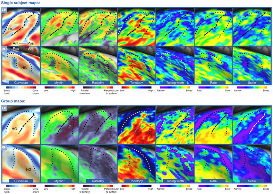
Fig. 3. Identification of PAC using various MR contrasts.
The upper and lower panel show an individual participant and group data (N = 10), respectively. The anteromedial part of Heschl’s gyrus (HG), putative PAC, is functionally characterized by a complete tonotopic gradient, narrow tuning width, fast temporal modulation rates, and broad spectral modulation scales. Myelination is high in this region, and radiality is greater (i.e., more perpendicular to the WM–GM surface) in the medial part of HG than the lateral part. The dashed and dotted black line identify the outline of HG and the intermediate sulcus, respectively. Figure from Gulban et al. (2017).

