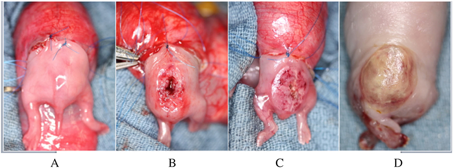
Figure 2:
Gross views of: A) the exposed hindlimbs and rump of a rabbit fetus prior to creation of a spina bifida defect; B) completed spina bifida defect; C) completed application of the composite adhesive system covering the defect; D) adherent composite covering the spina bifida defect after euthanasia.

