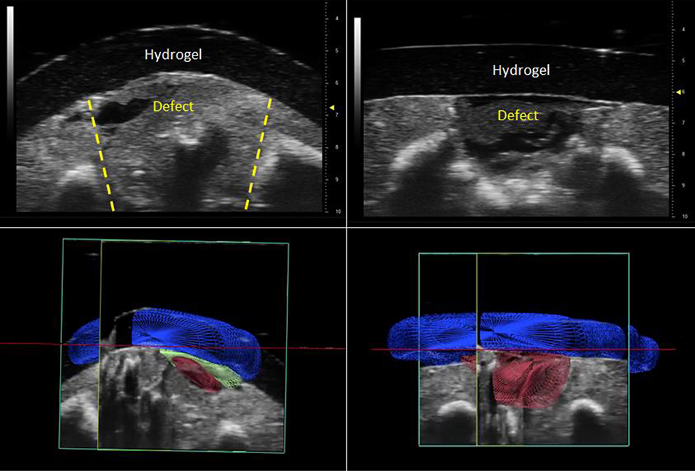
Figure 3:
High-frequency ultrasound was used to demonstrate adhesion between the hydrogel and fetal tissue in both operated (2D: upper left; 3D: lower left) and time-zero (2D: upper right, 3D: lower right) fetuses after euthanasia. In the 3D ultrasound images, 3D renderings of different structures were created based upon relative echogenicity as follows: the hypoechoic hydrogel is rendered in blue, the hypoechoic defect is rendered in red, and a layer of hyperechoic tissue interface between the hydrogel and defect is rendered in green.

