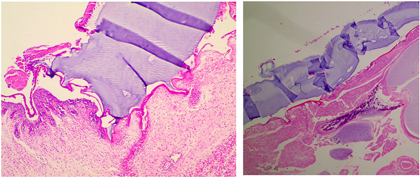
Figure 4:
Representative histological images showing: (left) integration of the hydrogel (purple), chitosan bioadhesive (bright pink), and fetal tissue, with interdigitation of the hydrogel-adhesive composite into the fetal tissue; (right) lack of adhesion of the hydrogel to the underlying flattened spinal cord typical of spina bifida. (H&E, 100x).

