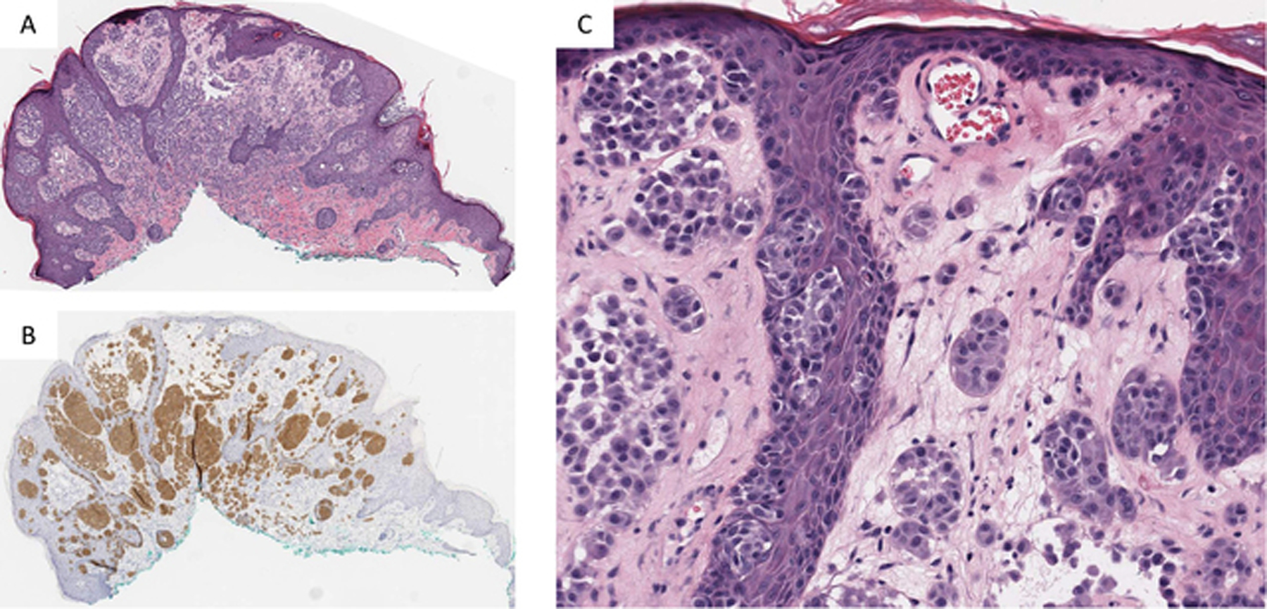
Figure 4).

A) Low power showing plaque like silhouette of a ROS1 Fusion Spitz nevus. B) IHC staining for ROS1 shows strong and uniform staining throughout the nevus. C) Higher magnification shows nests of epithelioid and spindle shaped melanocytes with bland cytomorphology lacking significant atypia.
