
Extended Data Fig. 6. Comparison of the nucleosome-bound ATPase domain of Snf2, RSC, and RSCSAR in the ADP-BeF3 nucleotide state.
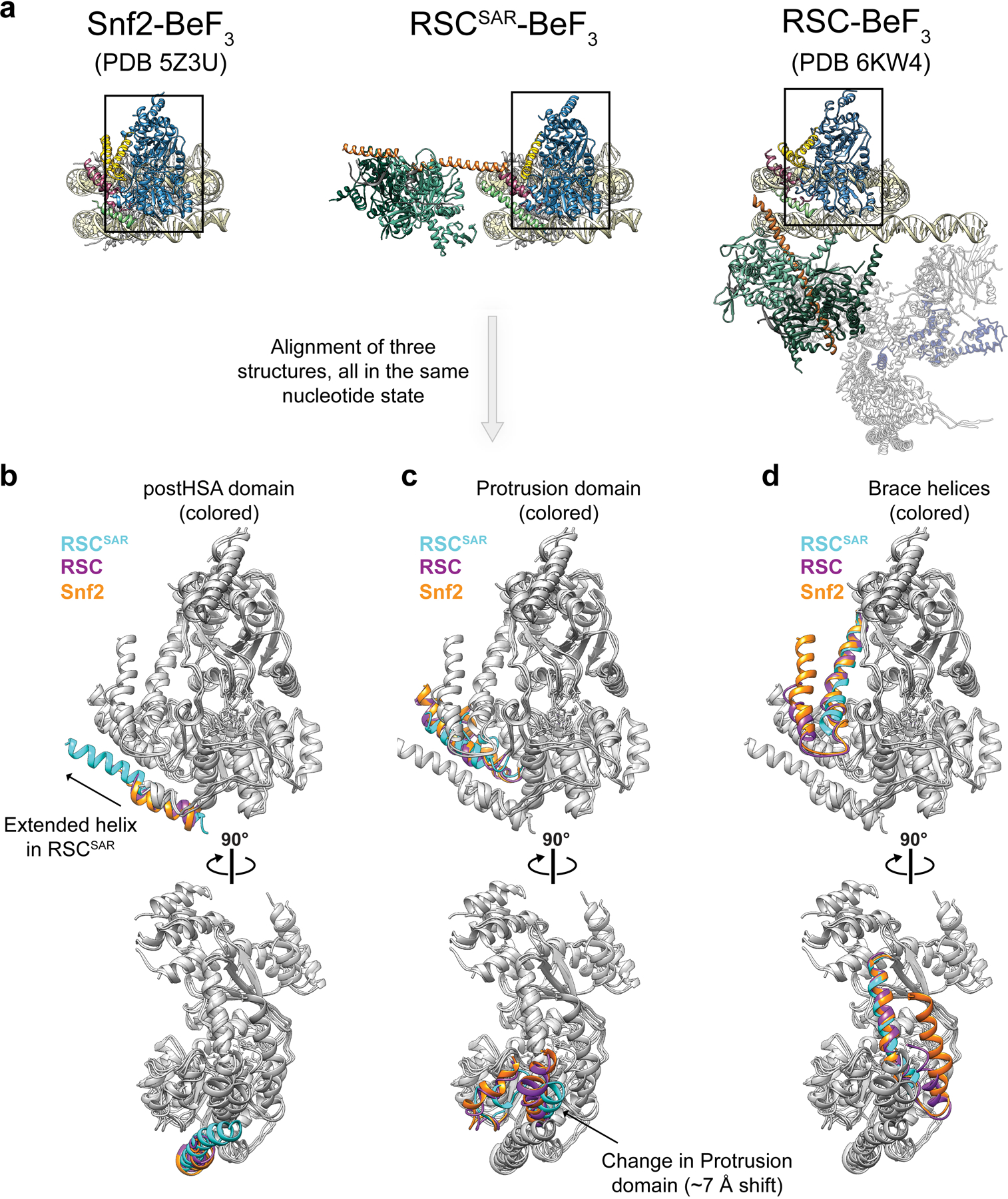
a, The PDB models for Snf2, RSCSAR, and RSC are shown with the same orientation and domain coloring. All three structures are from S.cerevisae, bound to the nucleosome and in the ADP-BeF3 nucleotide state. b-d, The ATPase domain of each structure from (a) was docked into the cryo-EM map of RSCSAR. The postHSA domain (b), Protrusion 1 domain (c), and Brace Helices (d) were colored according to their respective PDB model in each panel. Cyan: RSCSAR, Magenta: RSC (PDB 6KW3), Orange: Snf2 (PDB 5Z3U).