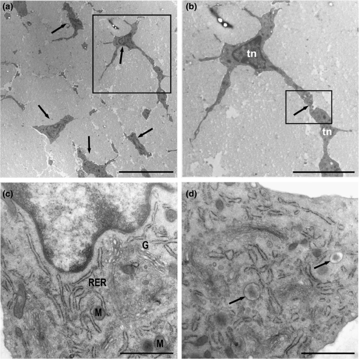
FIGURE 1.

Ultrathin sections of the equine digital flexor tendon. (a) Tendon areas showing polygonal and stellate tenocytes (arrows) immersed in a wide area of extracellular matrix. (b) Detail of A‐Tenocytes (Tn) with long slender cytoplasmic processes that contact neighbouring tenocytes (inset). (c) Tenocytes with rough endoplasmic reticulum (RER), a well‐developed Golgi apparatus (G) and few mitochondria (M). The nucleus contains a narrow layer of marginal chromatin. (d) A large number of vesicles released from the Golgi apparatus showing an electron‐dense central core containing matrix proteins were observed (arrows). Scale bars: (a) 10 µm; (b) 5 µm; (c) 1 µm; (d) 1 µm
