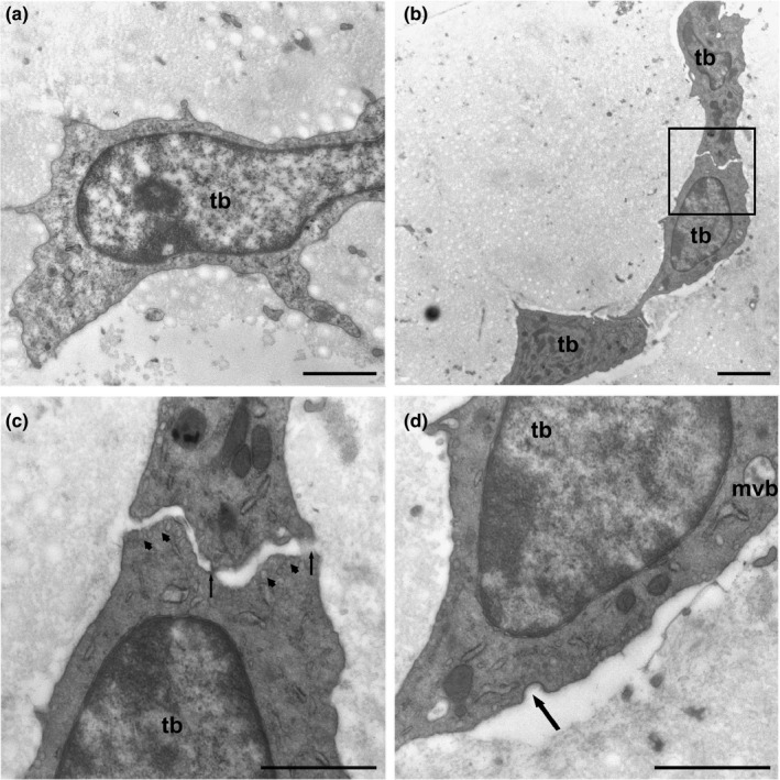
FIGURE 2.

(a) Tenoblast (tb) showing a voluminous nucleus and prominent nucleolus. (b) The cytoplasmic prolongations were wider and shorter than those in tenocytes and established contacts with neighbouring tenoblasts (inset). (c) Detail of B‐Tenoblast membranes made punctate contacts with electron‐dense reinforcements (arrows). Numerous pinocytic vesicles appear below the plasma membrane (arrowheads). (d) Tenoblasts (tb) have small saccules of rough endoplasmic reticulum and multivesicular bodies (mvb) containing exosomes. Numerous pinocytic vesicles and coated pits are indicative of endocytic processes mediated by clathrin (arrow). Scale bars: (a) 1 µm; (b) 2 µm; (c) 1 µm; (d) 1 µm
