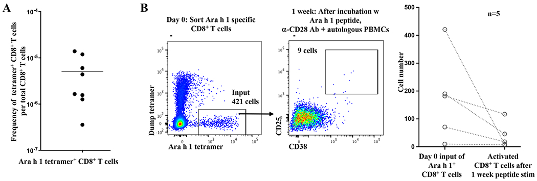
FIG 1.

A, Ara h 1 specific CD8+ T cells are not clonally deleted in nonallergic individuals.
Tetramer enrichment using HLA-A*02:01 tetramers loaded with Ara h 1 peptide was performed on PBMCs from HLA-A*02:01+ blood bank donors, after which peanut (Ara h 1) tetramer+ CD8+ T cells were enumerated by flow cytometry. Each point represents one blood bank sample.
B, Poor activation of Ara h 1 specific CD8+ T cells in nonallergic individuals.
Left: Ara h 1 specific CD8+ T cells were isolated from a HLA-A*02:01+ blood bank donor by tetramer enrichment followed by FACS. Ara h 1 specific CD8+ T cells were incubated one week with Ara h 1 peptide (1.5ug/ml), anti-CD28 antibody (5ug/ml), and autologous PBMCs as feeder cells before analysis. Flow cytometry panels are gated on CD8+ T cells.
Right: Cumulative results from 5 blood bank samples. Each pair of points connected by a line represents one sample. In some cases, FACS was not performed and instead 1/11th of the tetramer enriched cells was analyzed by flow cytometry to calculate the number of Ara h 1 specific CD8+ T cells, and the remainder used for tissue culture. The p value for a 2 tailed Wilcoxon signed rank test was 0.0625, which is the minimum value possible for n=5.
