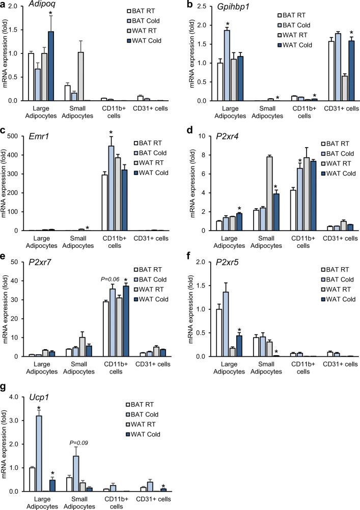
Fig. 2.
Gene expression analysis in cell fractions from adipose tissue (a–g). Male C57BL6/J WT mice were housed for 1 day at RT (22 °C) or cold (4 °C) condition. iBAT and subWAT tissues were harvested from each group, and different cell types were isolated by MACS®. Purity of cellular fractions was verified by gene expression of cell-specific markers for adipocytes (Adipoq), endothelial cells (Gpihbp1), and tissue resident macrophages (Emr1). The expression of P2rx4, P2rx5, P2rx7, and Ucp1 was determined in all isolated cell fractions. n = 4. Statistical analysis was done between cells of RT and cold group isolated from each tissue. Data are presented as mean ± SEM, *P < 0.05

