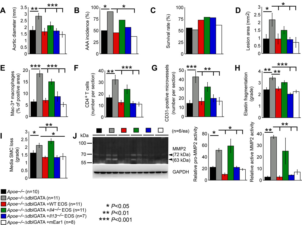
Figure 6.
EOS-derived IL4 and mEar1 are essential to Ang-II perfusion-induced AAA. Apoe–/–ΔdblGATA mice received adoptive transfer of EOS from WT, Il4–/– and Il13–/– mice, or osmatic minipump delivery of mEar1 followed by Ang-II infusion-induced AAA for 28 days. A. Abdominal aortic diameter. B. AAA incidence rate. C. Survival rate after Ang-II perfusion. D. AAA lesion area. E. Lesion Mac-3+ macrophage-positive area. F. Lesion CD4+ T-cell number. G. Lesion CD31+ microvessel number. H. Lesion elastin fragmentation grade. I. Lesion media SMC loss grade. J. Gelatin gel zymography detected lesion MMP2 and pro-MMP2 activity. Representative blot is shown to the left. The number of experiments and the number, genotype, and treatment of each cohort of mice are indicated. Data are mean±SEM. *P<0.05, **P<0.01, ***P<0.001, one-way ANOVA test.

