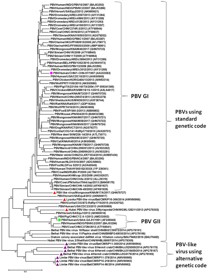
Figure 2.
Phylogenetic analysis of the picobirnavirus (PBV) and PBV-like RNA-dependent RNA polymerase (RdRp) sequences. The phylogenetic tree was constructed by the Maximum Likelihood method using the MEGA6 software. Phylogenetic distances were measured using the LG + G model of substitution. The tree was statistically supported by bootstrapping with 500 replicates. Bootstrap values <70% are not shown. Scale bar, 0.5 substitutions per amino acid. The name of the PBV strain includes virus/host of detection/country/common name/date of collection. GenBank accession numbers are shown in parentheses. Pink circle: prototype PBV genogroup-I (GI) strain; green circle: prototype PBV GII strain; purple triangles: PBV-like viruses that use an alternative mitochondrial genetic code to translate the RdRp and cluster separately from PBVs using the standard genetic code; red triangles: PBV-like strains that use an alternative mitochondrial genetic code to translate the RdRp, yet cluster within PBVs using standard genetic code.

