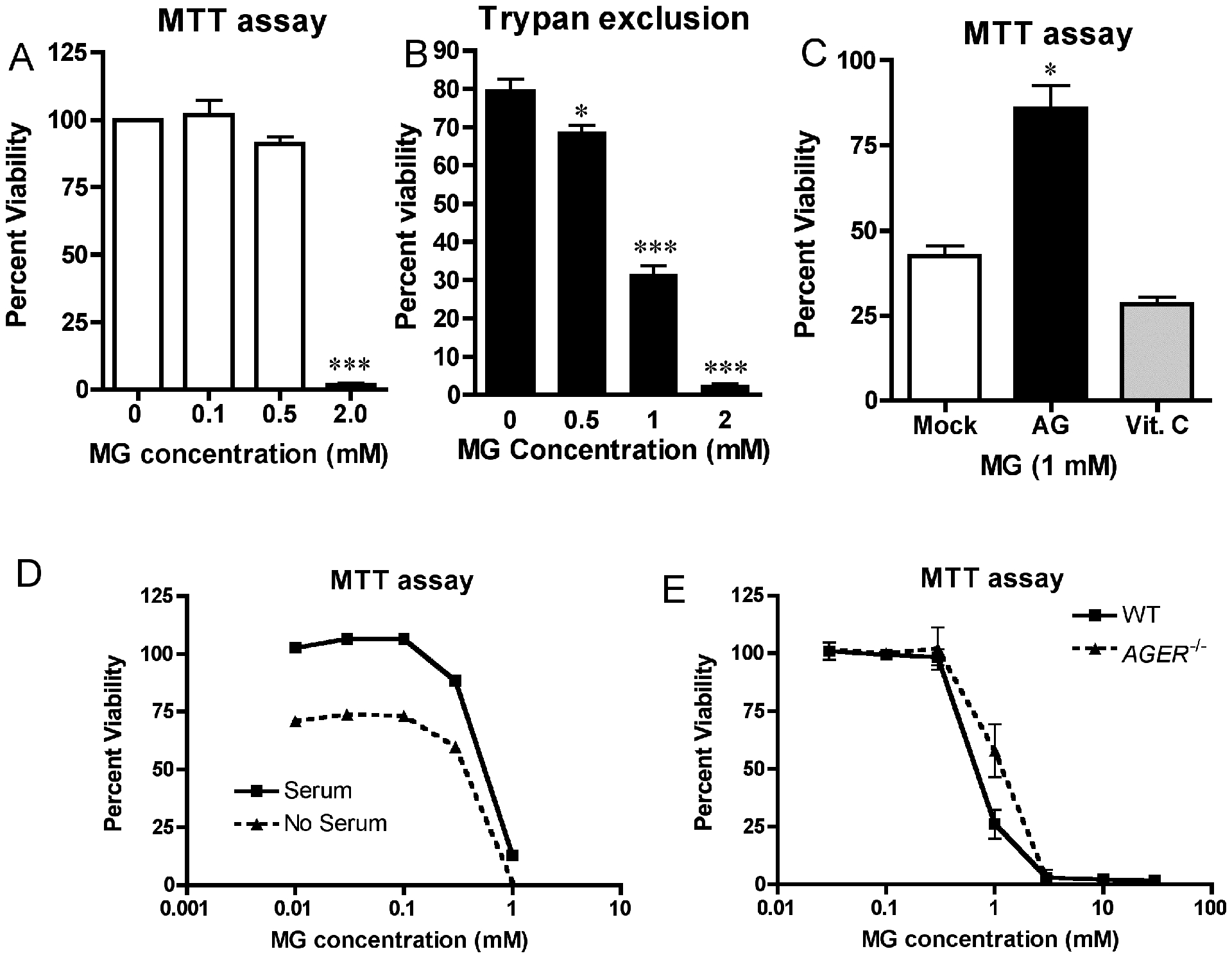
Figure 4: Cell death induced by MG is reversible by pretreatment with AG, but is RAGE-independent.

WT macrophages were stimulated in duplicate with the indicated doses of purified MG. Cellular viability was determined after 24 hours of incubation by Mitochondrial reductase (MTT) activity assay (A) or by trypan exclusion assay (B). (C) WT macrophages were treated as described in (A), except cells were incubated in the presence of the MG quencher, AG (1 mM), or the antioxidant, Vitamin C (5 mM), for 30 min prior to MG exposure. MTT activity was measured as in (A). (D) Macrophages were treated as in (A) except that serum was removed (or not) immediately prior to MG exposure. (E) WT and Ager−/− macrophages were stimulated with the indicated doses of MG. MTT activity was determined after 24 hours of incubation. Data for all panels represents the percent viability (mean ± SD) compared to untreated control cells and represent the results of 3 independent experiments. * Denotes p < 0.05. *** denotes p < 0.001.
