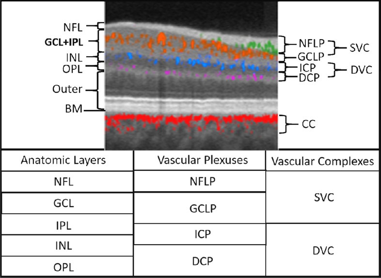
Figure 3.
Relationship between the retinal vascular plexuses and anatomic layers illustrated on a cross-sectional projection-resolved (PR) OCTA of a normal eye. Flow signals are color coded according to plexus and overlaid on a gray-scale reflectance image. Anatomic slabs (labels to the left of the image) are NFL = nerve fiber layer; GCL = ganglion cell complex, IPL = inner plexiform layer; INL = inner nuclear layer; OPL = outer plexiform layer; BM = Bruch’s membrane. Vascular plexuses (labels on the right of the image) are NFLP = nerve fiber layer plexus; GCLP = ganglion cell layer plexus; ICP = intermediate capillary plexus; DCP = deep capillary plexus. The GCLP extends from the NFL 67% of the way through the GCL and IPL. The vascular complexes are SVC = superficial vascular complex (NFLP + GCLP); DVC = deep vascular complex (ICP+DCP); CC = choriocapillaris. The table below gives the relative location of the retinal anatomic layers, vascular plexuses, and vascular complexes. Adapted with permission from (Campbell et al., 2017b).

