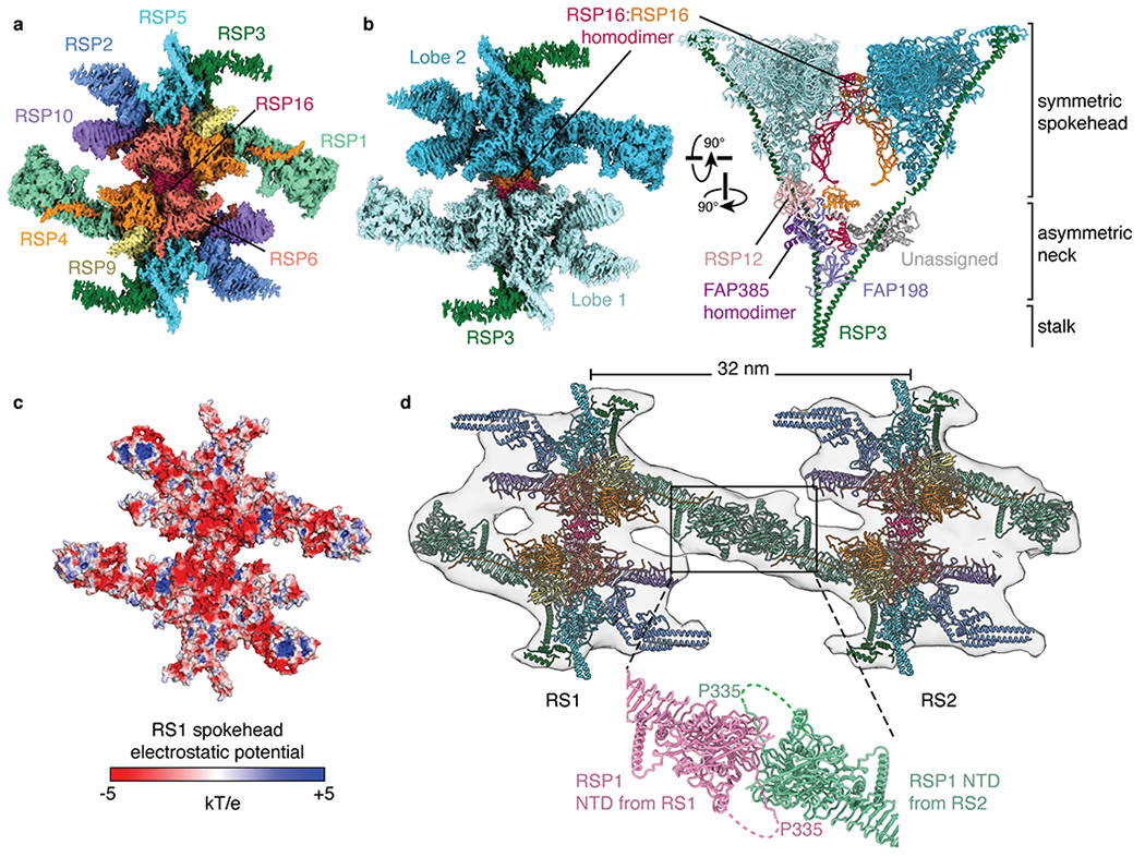
Fig. 3 |. Structure of the radial spokehead.

a, View of the top of the radial spokehead. Composite map colored by subunit. b, Two views of the radial spokehead. The two symmetric lobes of the spokehead (colored different shades of blue) are dimerized by a homodimer of RSP16 and flanked by the α-helices of RSP3. The symmetric spokehead sits on a V-shaped asymmetric neck containing FAP198, FAP385, RSP12, and the N-terminal domains of RSP16. c, Atomic model of the radial spokehead colored by electrostatic potential. d, Atomic models of the radial spokes docked into an isosurface rendering of the subtomogram average of the Chlamydomonas axoneme (EMD-6872). The two radial spokes interact through the N-terminal domains (NTD) of RSP1. The zoom-in shows the model of the on-doublet interaction between the NTDs of RSP1 from RS1 and RS2.
