
Fig. 2.
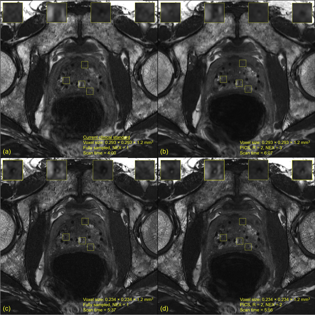
Magnetic resonance imaging (MRI) of the same patient imaged with multiple in-plane spatial resolutions and accelerated signal averages. Supplementary Figure E12 compares 1 strand in a sagittal view in normal magnification across the 4 acquisitions. (A) Fully sampled constructive interference in steady-state (CISS) MRI acquired at the moderate in-plane spatial resolution, which is the current clinical standard. (B) Accelerated CISS MRI acquired at the moderate in-plane spatial resolution, R = 2, number of signal averages = 3, and reconstructed with parallel imaging and compressed sensing. A notable increase in signal to noise was achieved compared with (A). (C) Fully sampled CISS MRI acquired at the high in-plane spatial resolution. Increased visualization of the implanted seed markers was achieved compared with that in (A). (D) Accelerated CISS MRI acquired at the high in-plane spatial resolution, R = 2, number of signal averages = 2, and reconstructed with parallel imaging and compressed sensing. An improvement in signal to noise was achieved compared with that in (C). These 4 MRIs are consecutive acquisitions of the same patient; because of the small voxel sizes, slight interscan patient motion may have caused minor shifts in anatomic landmarks, potentially causing slight differences in appearance of the seed markers.