Figure 1.

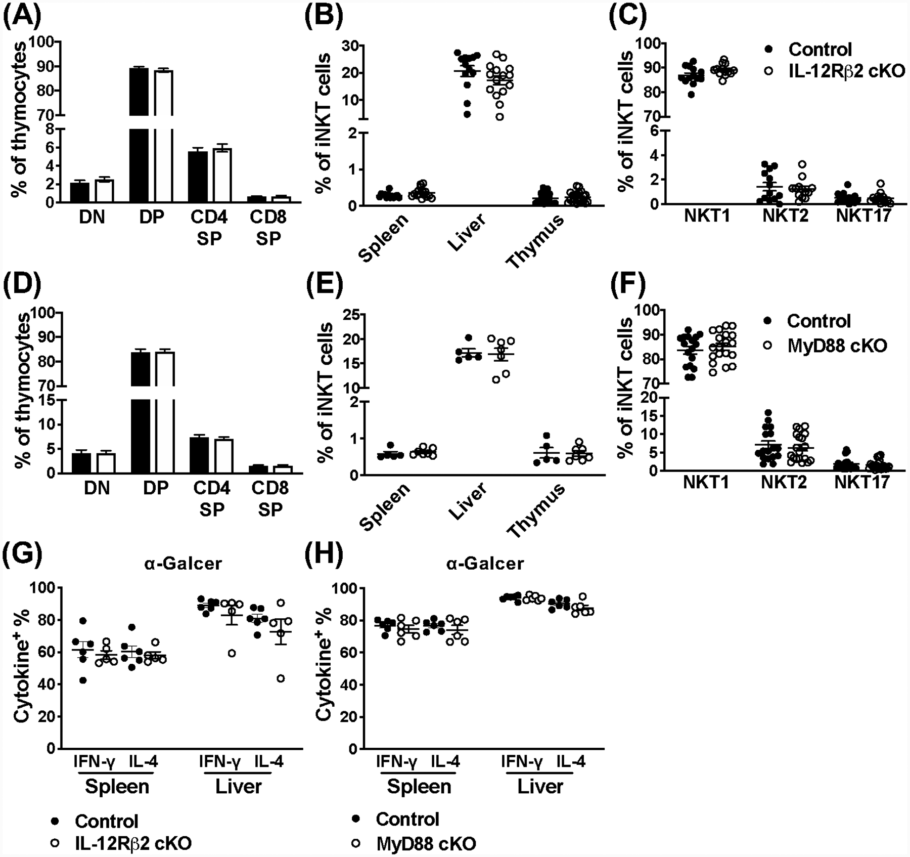
IL-12Rβ2 and MyD88 signaling are dispensable for iNKT cell development, peripheral localization, and their response to a strong agonist. (A) The CD4−CD8− double negative (DN), CD4+CD8+ double positive (DP), and CD4+ or CD8 + single positive (SP) stages of T cell development in the thymus of IL-12Rβ2 control (black) and cKO (open) mice (n=18–20). (B) Frequency of iNKT cells (TCRβ+CD1dtet+) in indicated organs from IL-12Rβ2 control (black) and cKO (open) mice (n=13–20). (C) Frequency of NKT1, NKT2, and NKT17 lineages from the thymus of IL-12Rβ2 control (black) and cKO (open) mice (n=13). NKT cell lineages (TCRβ+CD1dtet+) were differentiated using PLZF and RORγt expression. (D) The DN, DP, and CD4+ or CD8+ SP stages of T cell development in the thymus of MyD88 control (black) and cKO (open) mice (n=17). (E) Frequency of iNKT cells (CD45+TCRβ+CD1dtet+) in indicated organs from MyD88 control (black) and cKO (open) mice (n=5–7). (F) Frequency of NKT1, NKT2, and NKT17 lineages from the thymus of MyD88 control (black) and cKO (open) mice (n=17–19). NKT cell lineages (TCRβ+CD1dtet+) were differentiated using PLZF and RORγt expression. (G) Frequency of IFN-γ+ and IL-4+ iNKT cells (TCRβ+CD1dtet+) from the spleen and liver of IL-12Rβ2 control (black) and IL-12Rβ2 cKO (open) mice 2 hours post-stimulation with α-Galcer (n=5–6). (H) Frequency of IFN-γ+ and IL-4+ iNKT cells (TCRβ+CD1dtet+eYFP+) from the spleen and liver of MyD88 control (black) and MyD88 cKO (open) mice 2 hours post-stimulation with α-Galcer (n=6). Data are pooled from two (E, G, H) or at least three (A-D, F) independent experiments and error bars indicate SEM.
