
Figure 3.
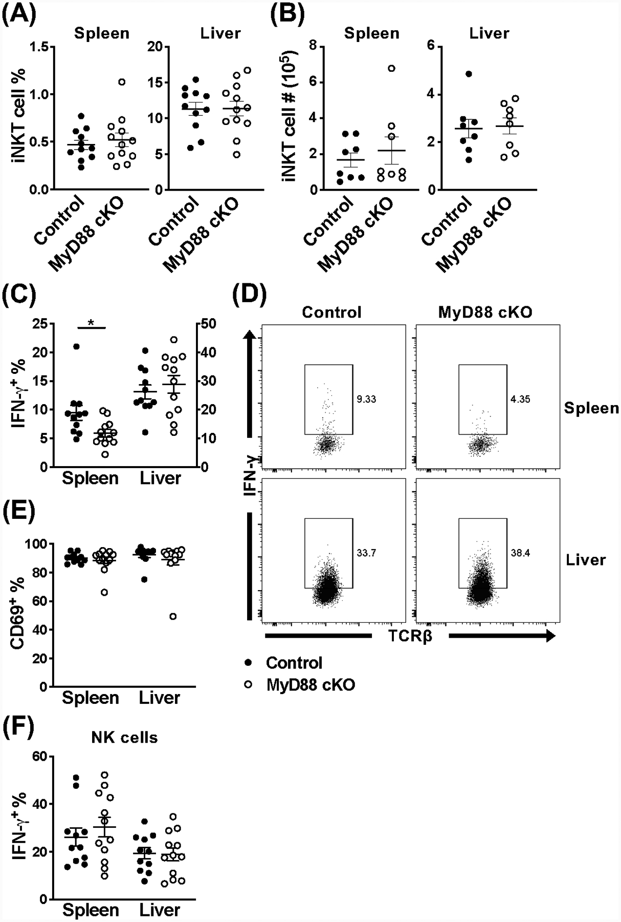
Splenic iNKT cells are hyporesponsive during MCMV infection in the absence of MyD88 signaling. (A) Frequency and (B) absolute number of iNKT cells (TCRβ+CD1dtet+) from the spleen and liver of MyD88 control (black) and MyD88 cKO (open) mice at 36 hours post-infection with MCMV. (C) Frequency and (D) representative flow cytometry of IFN-γ+ iNKT cells (TCRβ+CD1dtet+eYFP+) and (E) frequency of CD69+ iNKT cells (TCRβ+CD1dtet+eYFP+) from the spleen and liver of indicated mice at 36 hours post-MCMV. (F) Frequency of IFN-γ+ NK cells (TCRβ−NK1.1+) from the spleen and liver of indicated mice at 36 hours post-MCMV. Data are pooled from two (B, n=8) or three (A, C-F, n=11–12) independent experiments and error bars indicate SEM.