
Figure 3. Oxytocin Receptor (Oxtr) Expression and Responsiveness to Oxytocin in the Spinal GRP Neurons.
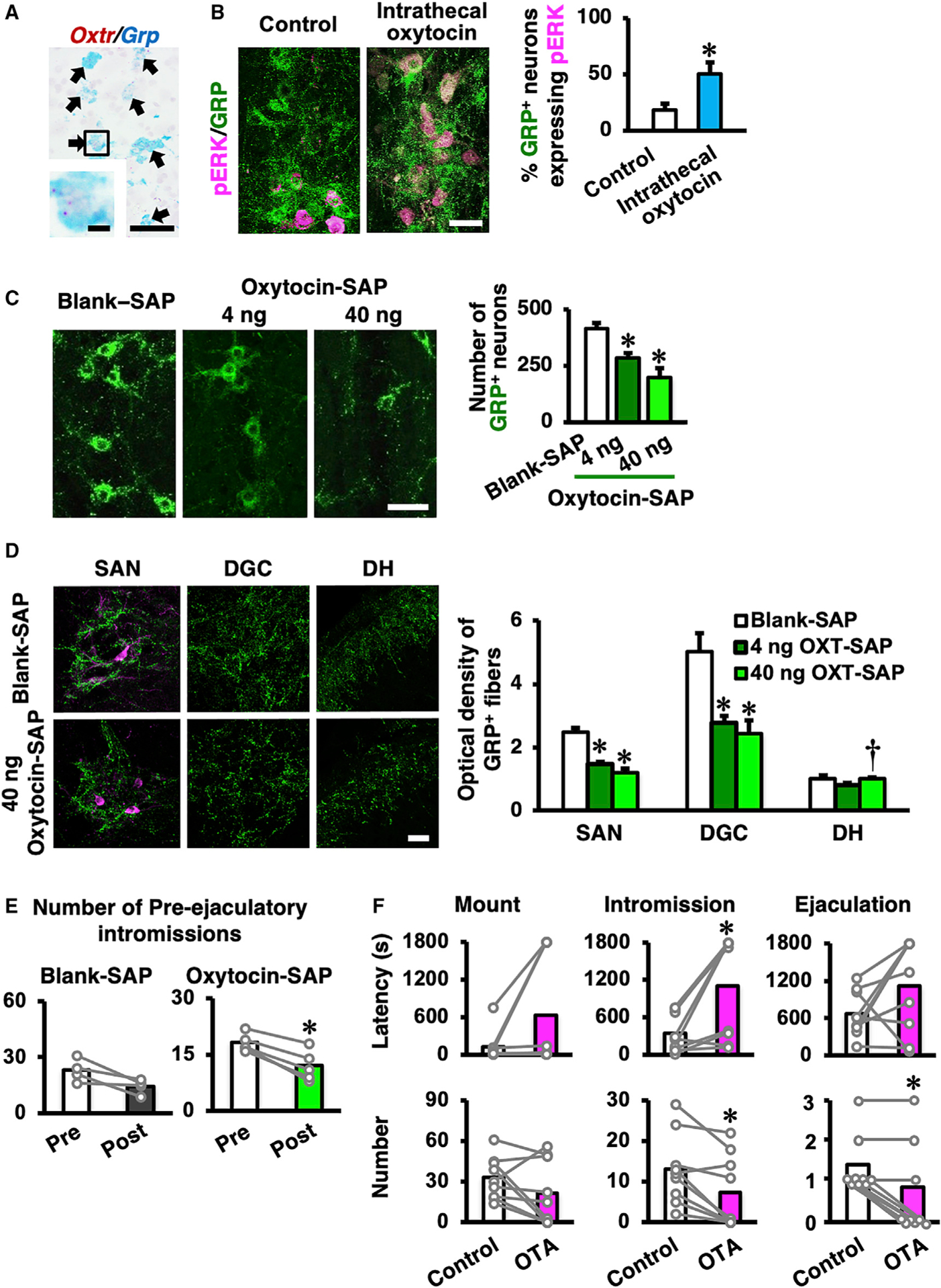
(A) Double in situ hybridization in male rats reveals that almost every Grp-positive neuron also expresses Oxtr mRNA. We could not find any Grp-positive but Oxtr-negative neurons in this study (n = 4, wild-type male rats). Scale bars: 50 μm (low magnification) and 10 μm (high magnification).
(B) Expression of pERK (magenta) in the spinal GRP neurons (green) after intrathecal oxytocin administration. Left panel: control group is shown (n = 4). Right panel: intrathecal oxytocin administration is shown (n = 5). Intrathecal oxytocin administration significantly increases pERK expression in GRP+ neurons compared to control (data are presented as mean ± SEM; Student’s unpaired t test; t6 = −2.74; *p < 0.05). Scale bar: 50 μm.
(C) The targeted toxin oxytocin-saporin (SAP), which consists of the toxin SAP conjugated to oxytocin was used. Oxytocin-SAP treatments (both 4 ng and 40 ng) significantly decrease the number of spinal GRP neurons (green) compared to the random peptide control, blank-SAP treatment. The left panel indicates blank-SAP-treated rats (n = 8). Middle panel: low-dose (4 ng) oxytocin-SAP-treated rats are shown (n = 7). Right panel: high-dose (40 ng) oxytocin-SAP-treated rats are shown (n = 6). Data are presented as mean ± SEM; one-way ANOVA; F2,18 = 15.2; *p < 0.05 versus blank-SAP. Scale bar: 50 μm. SEM, standard error of the mean.
(D) Optical density of GRP+ fibers in the lumbosacral spinal cord (L5–S1 level). Oxytocin-SAP-treated rats (4 ng, n = 7; 40 ng, n = 6) had fewer GRP+ fibers than did blank-SAP-treated rats (n = 8) in the sacral autonomic nucleus (SAN) and the dorsal gray commissure (DGC), but not in the dorsal horn (DH) (data as mean ± SEM one-way ANOVA; SAN, F(2, 18) = 20.5; DGC, F(2, 18) = 7.66; DH, F(2, 18) = 4.13; *p < 0.05 versus blank-SAP; †p < 0.05 versus 4 ng oxytocin-SAP). Scale bar: 50 μm.
(E) Effects of intraspinal administration of oxytocin-SAP (40 ng; n = 9) on male sexual behavior. Blank-SAP (n = 3) was used as control. The number of pre-ejaculatory intromissions of oxytocin-SAP (40 ng)-injected rats were decreased post-injection compared to pre-injection. No significant differences were observed in the other parameters of sexual behavior (data are presented as mean [highlighted] and individual dot [gray]; Student’s paired t test; pre-ejaculatory intromission number: blank-SAP t2 = 1.07; oxytocin-SAP t8 = 5.11; *p < 0.05).
(F) Effects of intrathecal administration of OXTR antagonist (OTA) on male sexual behavior. OTA intrathecal administration prolongs the latency to the first intromission and decreases the number of intromissions and ejaculations (data are presented as mean [highlighted] and individual dot [gray]; n = 9; Student’s paired t test; mount number: t8 = 1.63; intromission number: t8 = 3.43; ejaculation number: t8 = 3.16; *p < 0.05).