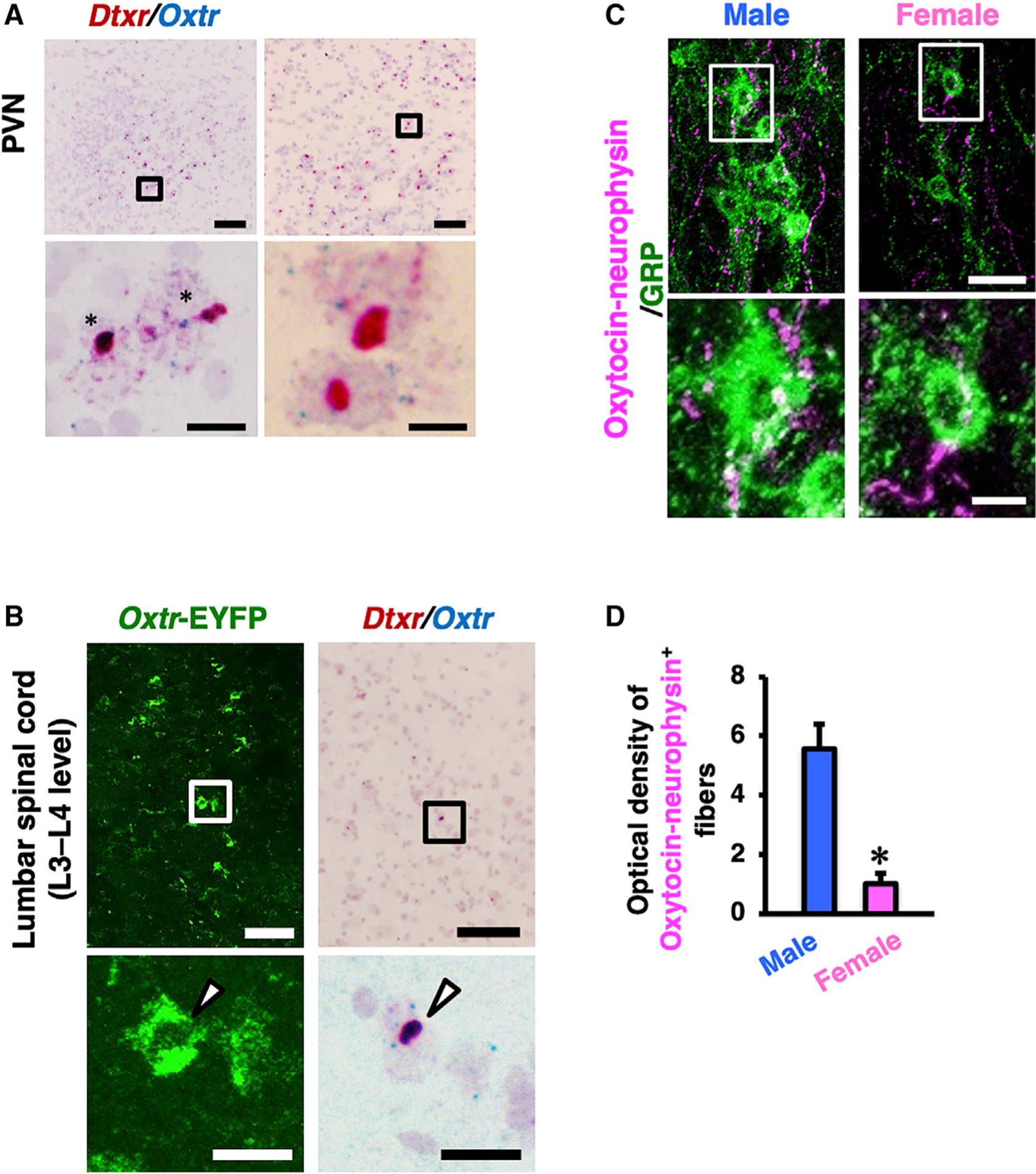
Figure 4. Characterization of Oxtr-CHR2-EYFP Transgenic Rats.

(A and B) Double in situ hybridization of human diphtheria toxin receptor (Dxtr) mRNA (red) and Oxtr mRNA (blue) in the PVN (A) and the lumbar spinal cord (L3 to L4 level; B). The lower panels are higher magnification images of the outlined areas in the upper panels. In the images of the PVN, almost all Oxtr+ neurons also express Dtxr (asterisks). In the images of the lumbar cord, arrow-heads indicate EYFP/Dxtr mRNA/Oxtr mRNA triple-positive neuronal somata. Scale bars: 100 μm (low magnification in A and B), 10 μm (high magnification in A), and 20 μm (high magnification in B).
(C) Double immunofluorescence for GRP (green) and oxytocin-neurophysin (magenta) in the lumbar cord of male and female rats demonstrated that oxytocin-containing axons surrounding GRP+ neurons exhibit a male-dominant sex difference.
(D) Quantitative analysis of oxytocin-neurophysin-immunoreactive axons in the lumbar spinal cord showed this sex difference (in the distribution—it is not really “distribution”). Outlined areas are enlarged in the lower panels. Data are presented as mean ± SEM. Student’s unpaired t test; oxytocin, t6 = 4.96; *p < 0.05; male rats (n = 4), female rats (n = 4). Scale bars: 50 μm in upper images and 20 μm in lower images.
See also Figures S2–S4.
