
Figure 6. Localized Volume Transmission by Exocytosis of Oxytocin in the Lumbar Spinal Cord Controls the Spinal Ejaculation Center.
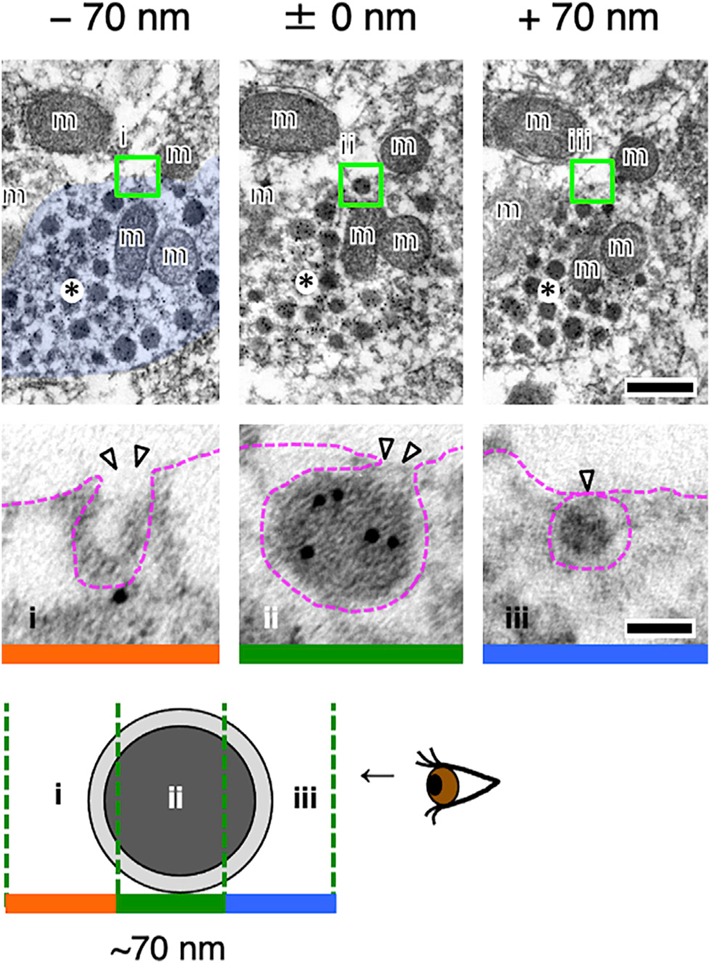
Three serial ultrastructural sections (~70 nm in thickness: i–iii) are displayed. Oxytocin-neurophysin is labeled with 10 nm gold particles. An oxytocin-neurophysin+ neurosecretory vesicle in the varicosity (asterisks) appears to be undergoing exocytosis at a non-synaptic site. Each blocked area is enlarged below, respectively. Arrowheads indicate exocytosis (i and ii) or docking (iii). m, mitochondrion. Scale bars: 200 nm in upper images and 50 nm in lower images. See also Figure S7.