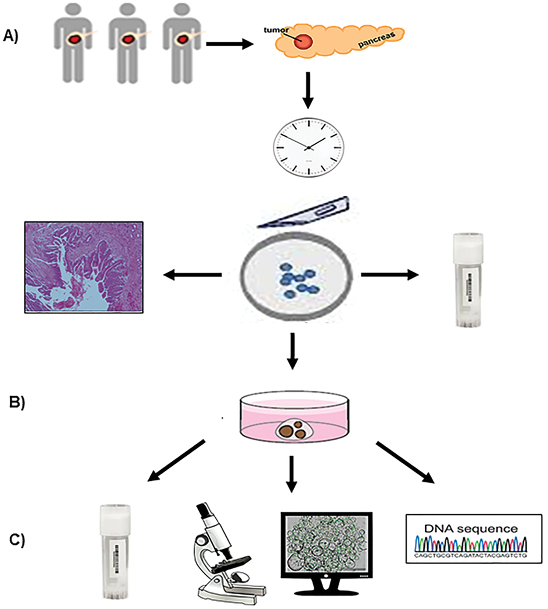
Figure 1. Workflow of patient-derived IPMN organoid culture methodology.

A) Resected IPMN tumor and adjacent normal tissues obtained from patients were transported to the processing lab within 20 minutes of resection; and a portion of each type of tissue was cryopreserved. Primary tissue was analyzed by histomorphology and immunohistochemistry analysis. Both IPMN tumor and adjacent normal tissue specimens were minced into small pieces and subsequently subjected to organoid culture. B) 3D-organoid cultures from both tumor and normal tissues were established. C) Organoids were cryopreserved and culture was imaged via bright-field microscopy, automated counting was conducted via the Circular Hough Transform-based algorithm, and characterized by genomic DNA fingerprinting and sequencing.
