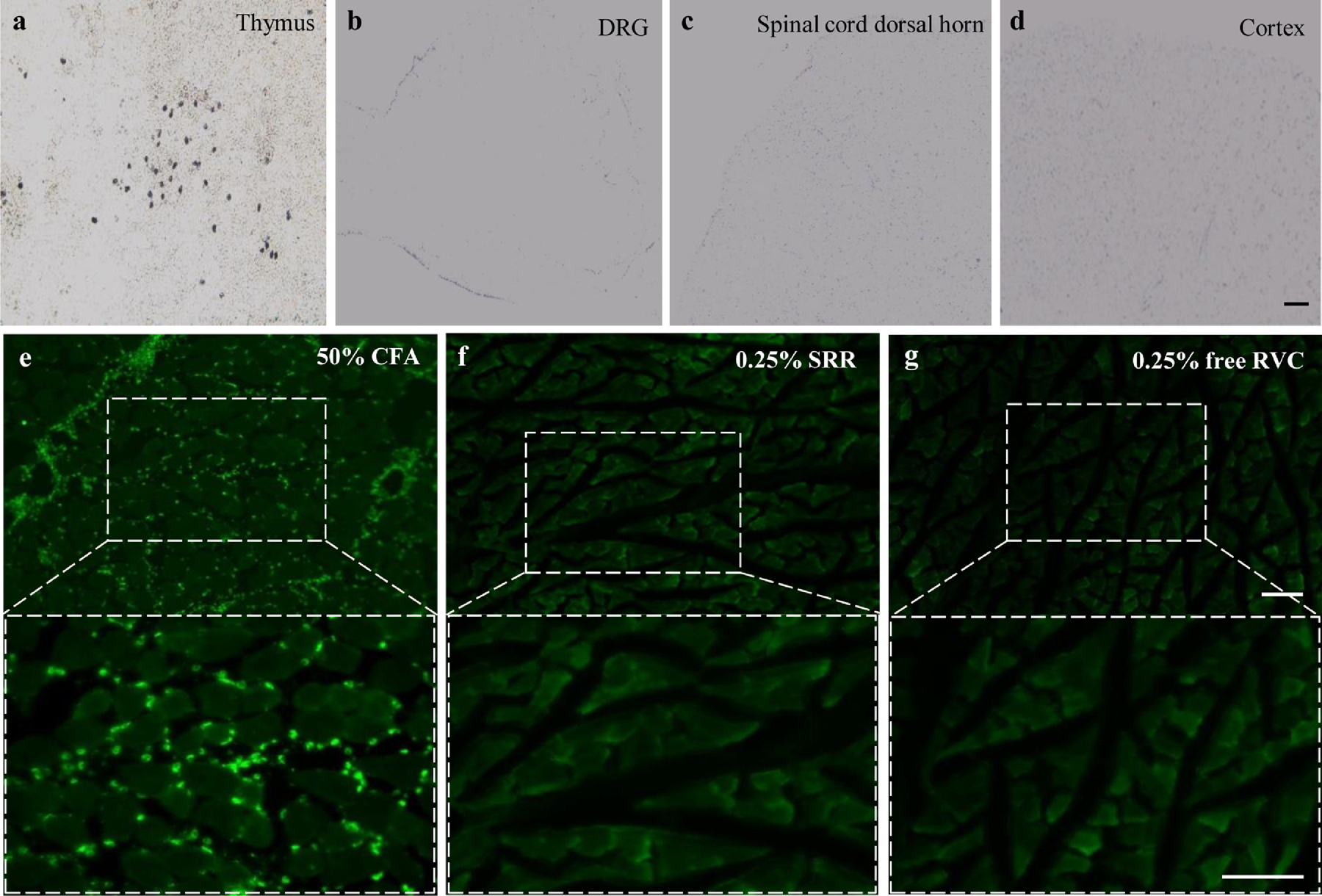
Figure 8.

(a-d) Representative photographs showing TUNEL-positive cells in the thymus (a), the fourth lumbar dorsal root ganglion (b), the fourth lumbar spinal cord dorsal horn (c) and brain cortex (d) on the ipsilateral side on day 15 after the peri-sciatic nerve injection of 0.25% SRR. n = 3 rats (15 sections)/group. Scale bar: 100 μm. (e-g) Representative images of CD68-like immunoreactivities in the muscles on day 15 after 50% complete Freund’s adjuvant (CFA) injection (e) and at the injected site after peri-sciatic nerve injection of 0.25% SRR (f) or 0.25% free RVC (g). n = 3 rats (15 sections)/group. Scale bar: 10 μm.
