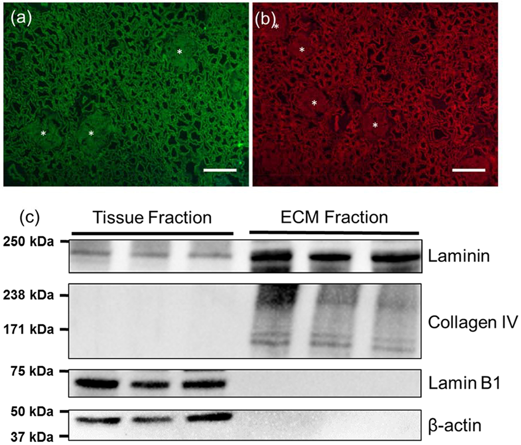
Figure 1.

Characterization of decellularized kidney cortex. (a,b) Collagen IV and laminin immunofluorescence images from decellularized cortex. Matrix showed minimal residual nuclear material based on counterstaining with DAPI. Scale bar=100 μm (c) Western blot analysis of laminin, collagen IV showing that structural ECM proteins are retained in the matrix following decellularization. Some laminin was extracted in the tissue fraction but was largely retained in the insoluble ECM fraction. Nuclear (Lamin B1) and cytoskeletal (β-actin) proteins were found in the tissue fraction but were not detected in the ECM fraction.
