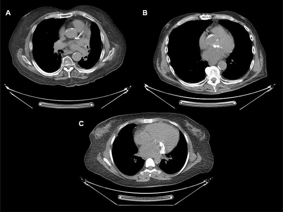
Figure 2. Attenuation correction computed tomography axial images from three patients with prior thoracic irradiation undergoing positron emission tomography/computed tomography myocardial perfusion imaging.
A) Extensive atherosclerotic calcification of left main artery, and proximal left anterior descending and left circumflex arteries, in addition to calcification of the aortic root and descending thoracic aorta. B) Extensive atherosclerotic calcification of the aortic root is evident on this ungated attenuation correction computed tomography axial image. C) Prominent mitral annular calcification is present.

