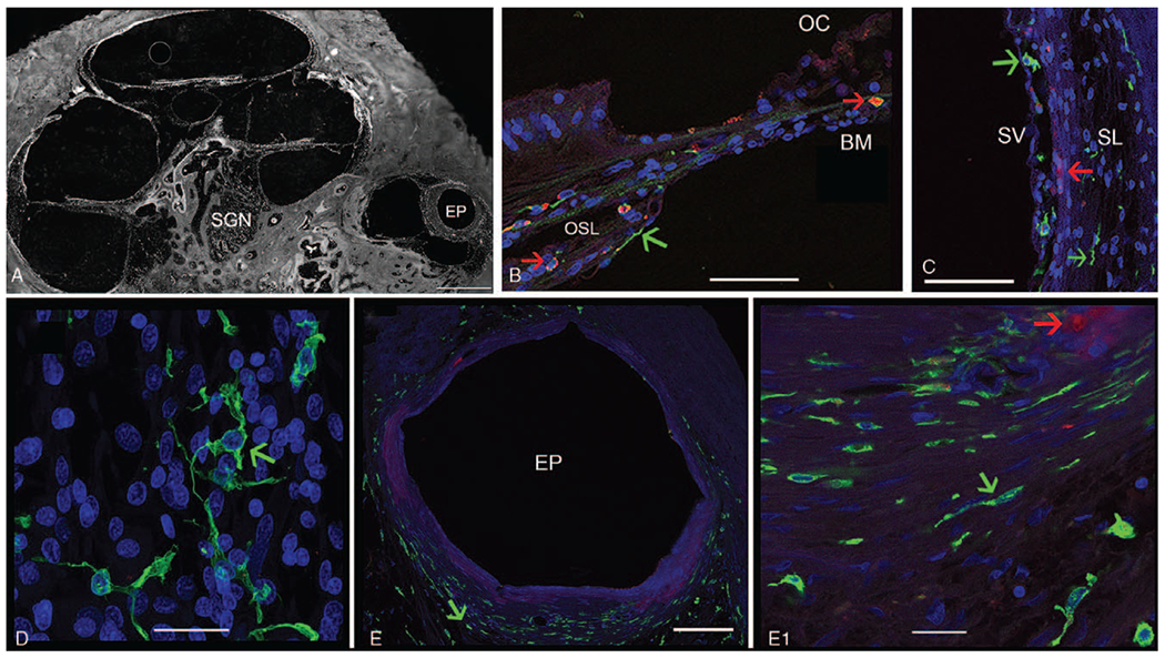
FIG. 1.

Immunofluorescence (IF) for Iba1 (green color) and CD68 (red color) in a CI cochlea. (Seventy-three-year-old female with a history of CI for 3 yr). A, Low magnification view of the implanted cochlea. EP indicates electrode path can be seen within the scala tympani; SGN, spiral ganglia neurons. B, Iba1-IF and CD68-IF macrophages are seen in the osseous spiral lamina (OSL) and basilar membrane (BM) underlying the organ of Corti (OC), with evidence of colocalization. C, IF macrophages in the stria vascularis (SV) and spiral ligament (SL). D, Iba1-IF macrophages in the spiral ganglia, exhibiting a ramified morphology E, Fibrous tissue encapsulation around the electrode path (EP) in the base-hook region of the cochlea (from (A)). Iba1-IF macrophages of elongated shape (green color) are seen surrounding the fibrous encapsulation. (E1) High magnification view from E, showing Iba1-IF macrophages (green short arrow), CD68-IF cells macrophages are visualized in red IF with a round morphology, and foamy appearance (red short arrow). DAPI indicates delineates cell nuclei. Counterstaining with Alcian blue shows the bony labyrinth. Bar in A= is 500 μm, E= 100 μm, E1= 20 μm, B to D= 40 μm.
